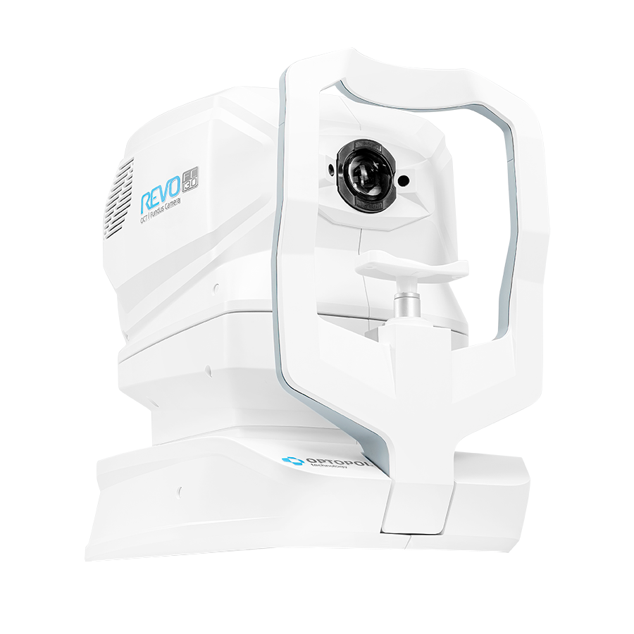
Take your OCT to the next level
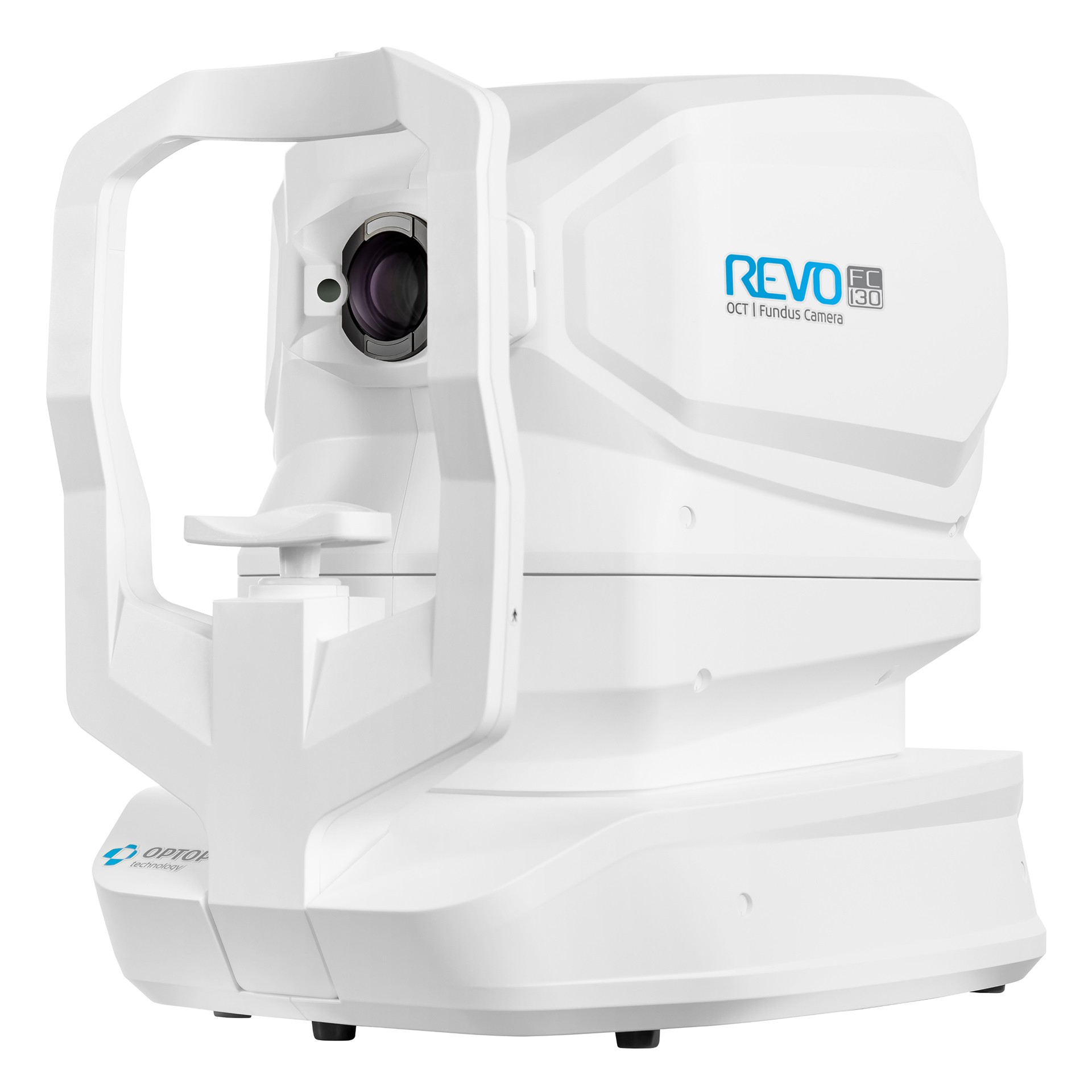
REVO FC 130
Auto Functions
Simplifying operation with the push of a button to auto-postion, auto-align, auto-focus, and auto-capture.
AccuTrack™
Our hardware-based eye tracker, compensates for blinks, loss of fixation and involuntary eye movements during scans reducing artifacts.

Full range
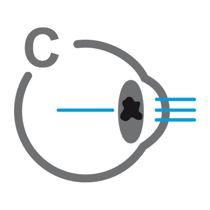
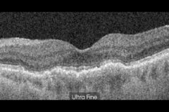
With scans presenting New Extended Depth™ software, based on our Full Range technology, this new imaging mode provides scans of increased depth for reliable and convenient observation of challenging cases. The Full Range mode is perfect for diagnosing even highly myopic patients.
Custom Scan Protocols
Save time and never miss a scan. Combine any scan type into a pre-set group. Choose a group of scans and set the order, the REVO will do the rest.

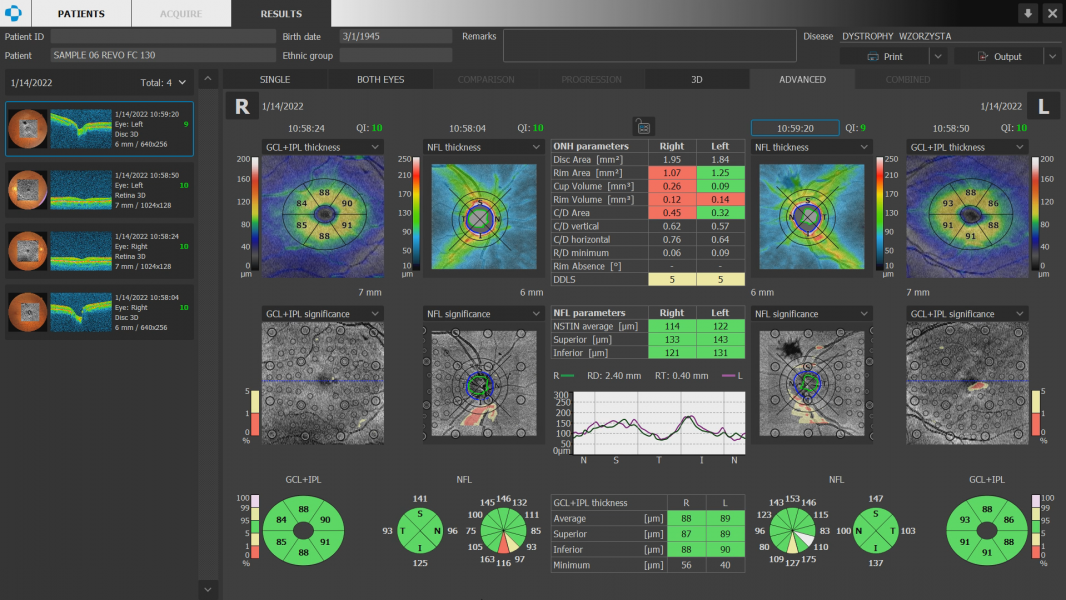
Structure&Function (S+F)
Comprehensive glaucoma solution that combines REVO OCT and PTS Visual Field results. S+F takes the diagnostic approach of the Hood report.Our S+F report allows clinicians to understand the relationship between structural glaucoma damage and the functional impact on the patient’s field of vision. This provides a quick and comprehensive single-page report for glaucoma management.

Glaucoma Tool Kit
Comprehensive glaucoma analytical tools for quantification of the Nerve Fiber Layer and Ganglion Cell Layer. The Disc Damage Likelihood Scale enables clinicians to precisely diagnose and monitor glaucoma for Optic Nerve Head analysis.
Progression Analysis
Quickly view a chronological set of exams for analysis of changes in morphology, quantified progression maps, and progression trends.

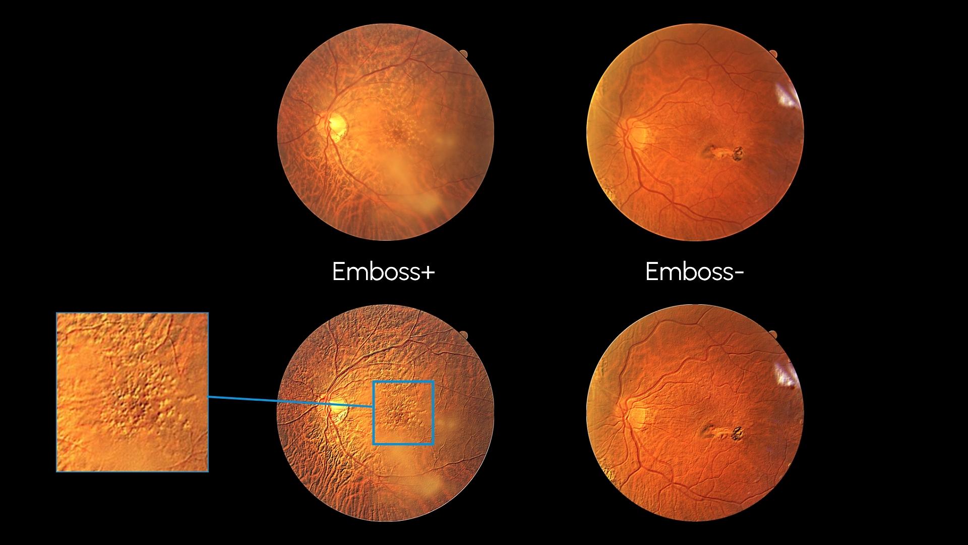
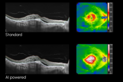
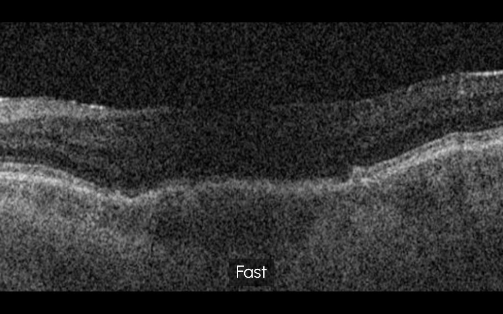
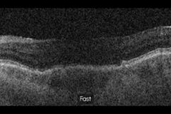
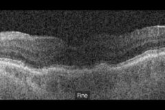
AI DeNoise
An advanced artificial intelligence (AI) algorithm removes noise from the tomogram for the highest image quality.
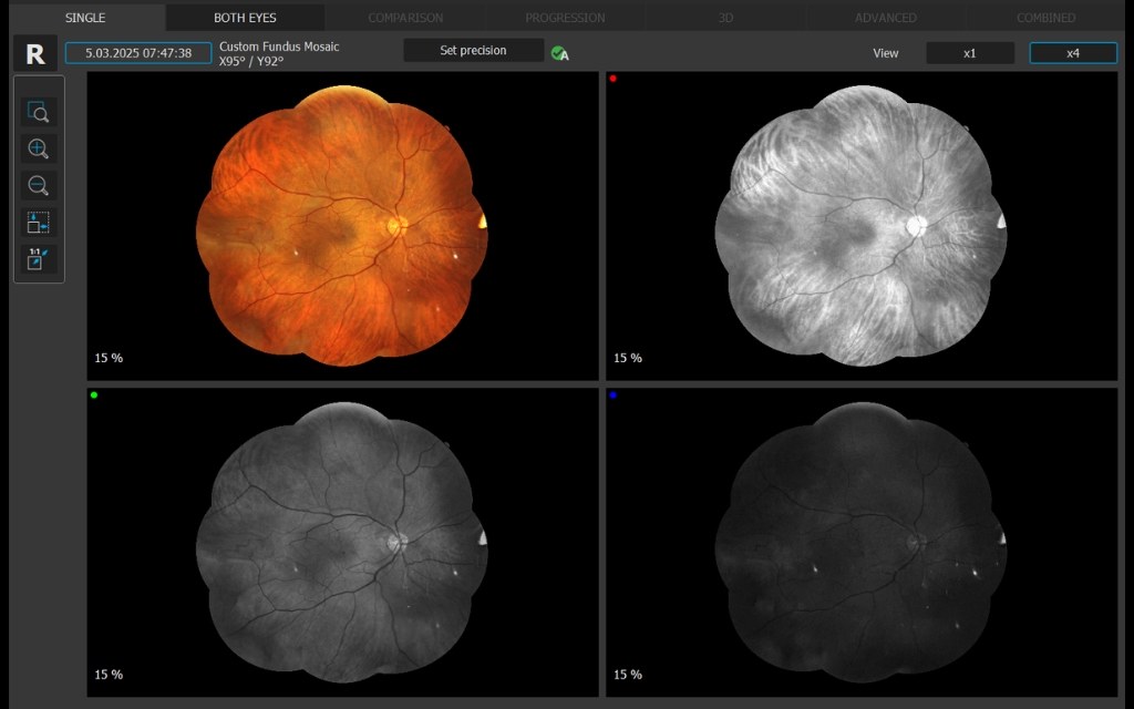
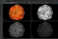
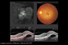
Non-Myd Color Fundus Camera
Integrated fundus and anterior imaging within a single OCT workflow
The REVO FC OCT series integrates a high-resolution 12.3 MP non-mydriatic fundus camera, enabling the capture of true-color retinal images with a 45° field of view (67.5° internal angle), delivering excellent detail and consistency. Fully automated operation — including auto-alignment, auto-focus, auto-flash, and auto-capture — ensures fast and user-independent image acquisition, even with pupil sizes as small as 3.3 mm.
The system also supports anterior segment photography, allowing documentation of corneal and anterior eye structures within the same workflow.

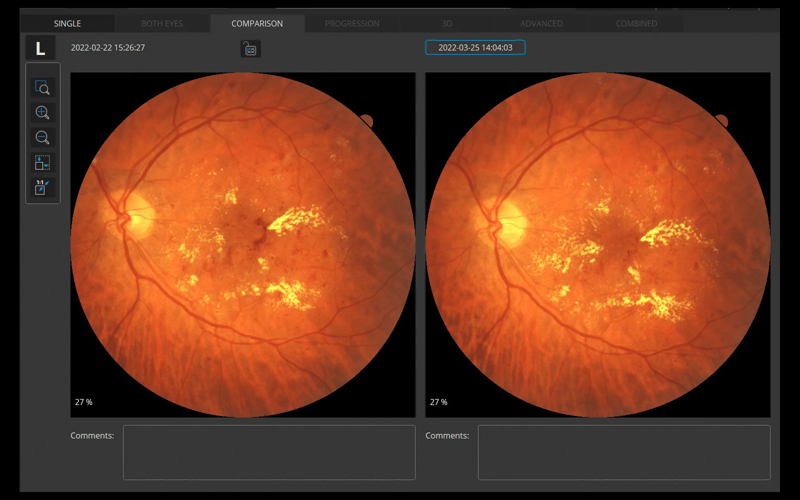
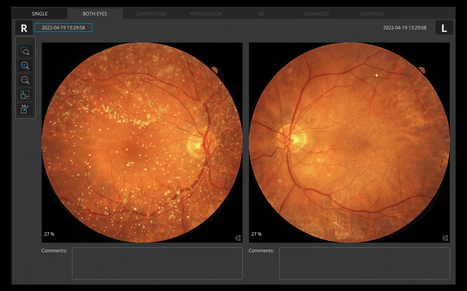
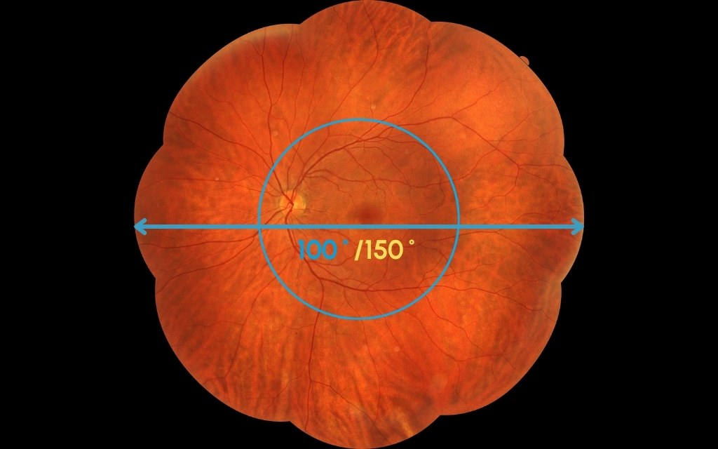
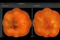
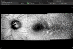
Panoramic Wide-Field Photography
REVO OCT devices with integrated fundus camera enable wide-field retinal imaging with coverage of up to 100° (~150° at the spherical center of the eye). In Mosaic photo mode, up to 9 internal fixation positions allow capture of peripheral areas and create a wide-field mosaic image — without mandatory patient dilation.
The REVO FC now enables coverage of up to 100° (~150° at the spherical center of the eye), providing a new wide perspective in retinography. In Mosaic photo mode, up to 9 internal fixation positions are available to capture peripheral areas and create a wide-field mosaic image. Patient dilation is not mandatory.
Fully automatic acquisition provides true-color fundus images in five configurations:
- Horizontal Fundus Mosaic x3
- Vertical Fundus Mosaic x3
- Fundus Mosaic x5
- Fundus Mosaic x9
- Custom Mosaic x2–9
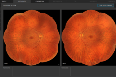
New EyeTracking
The updated EyeTracking function improves scan stability and tracking accuracy during image acquisition.
It ensures stable performance in eyes with darker retinal pigmentation (e.g. Asian population) and reduces dependency on pupil size conditions. The system also delivers improved tracking results with no delay in acquisition.*

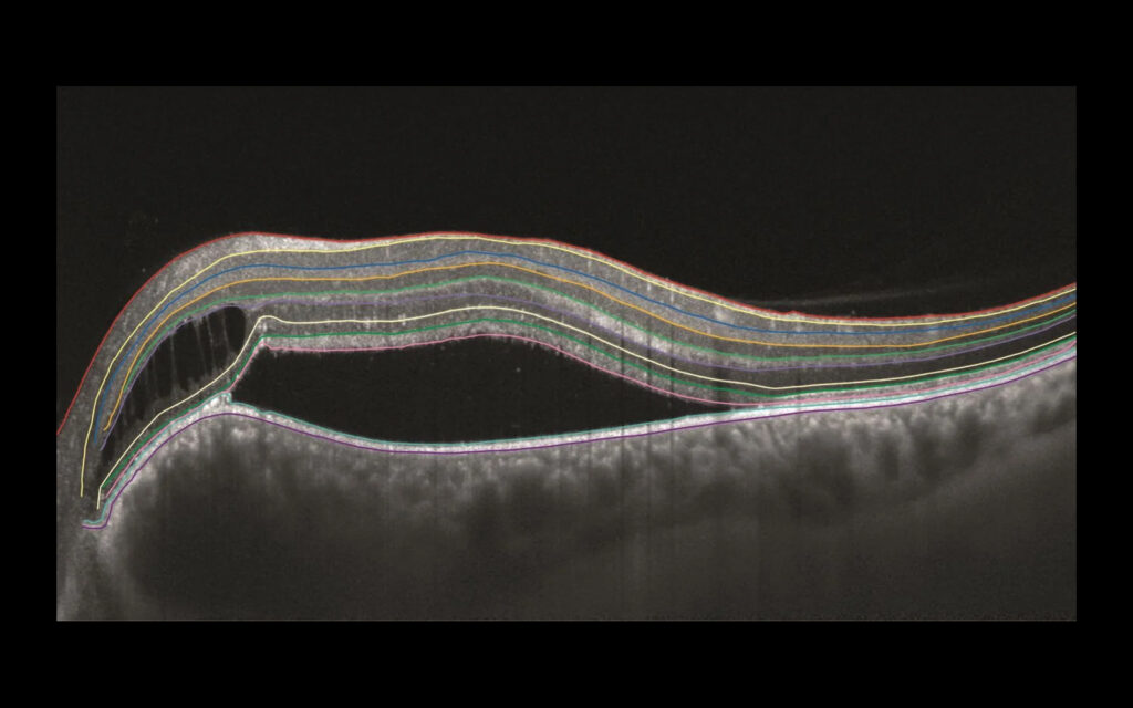
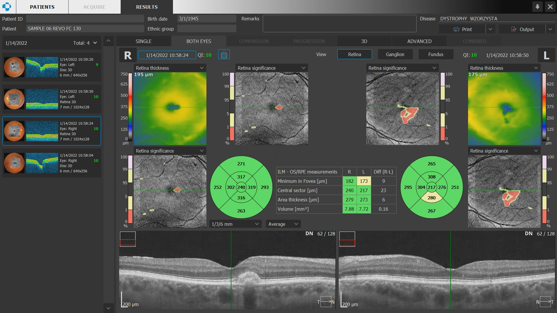
AI Posterior Segmentation
AI Retina
AI-based segmentation of the posterior segment enables automatic detection and analysis of up to 10 retinal layers, resulting in more accurate recognition of retinal layer boundaries. The AI system supports precise structural assessment and improves evaluation of pathological changes within the retina.


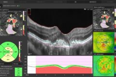
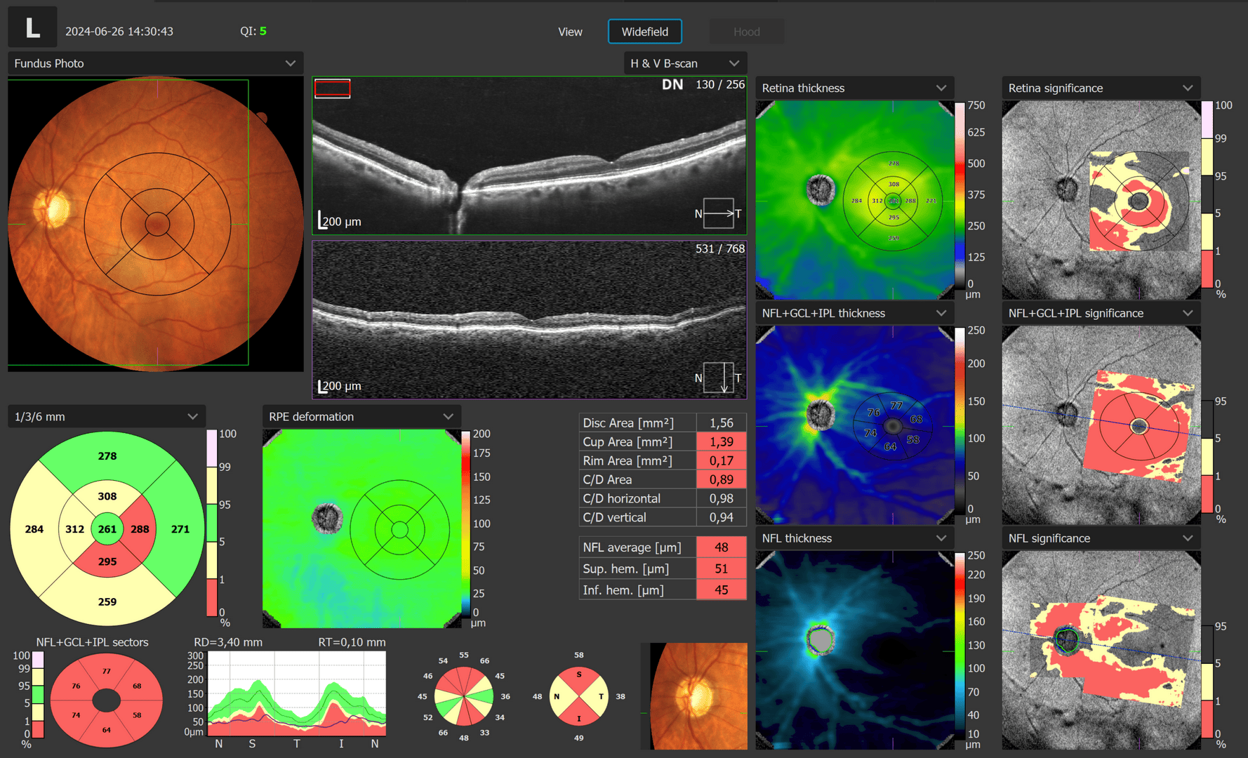
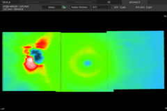
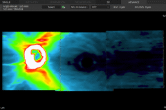
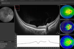
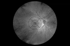
Widefield Analysis
A single Widefield 3D examination is now sufficient for the rapid assessment of both the retina and the optic nerve head, reducing examination time and eliminating the need for multiple separate scans.
Visualize and assess the thickness of the retina, ganglion cell layer, nerve fiber layer, and optic nerve head on a comprehensive data report when performing widefield mapping up to 15 × 15 mm.
The widefield report presents horizontal and vertical tomograms and includes optic disc topography, supporting efficient evaluation and follow-up of glaucoma patients.
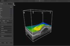
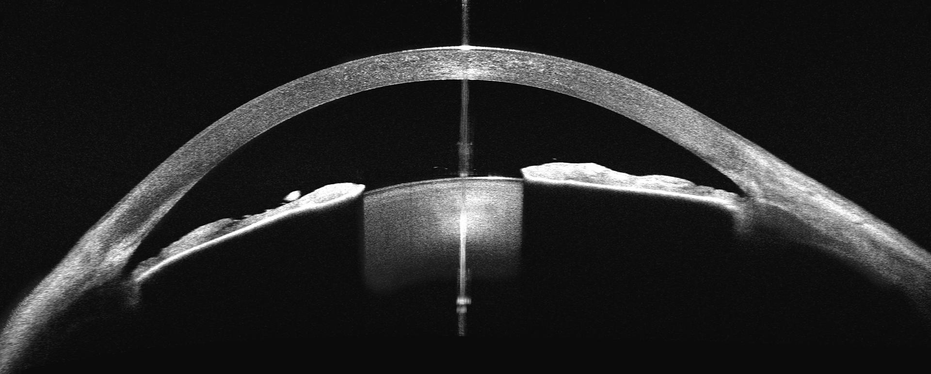
Anterior Chamber
Dedicated anterior segment protocols, combined with Full Range technology, enable detailed assessment of the anterior chamber, including visualization of the iridocorneal angle and measurement of anterior chamber depth.
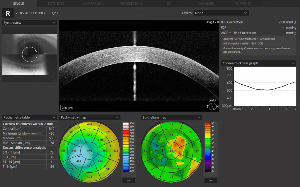
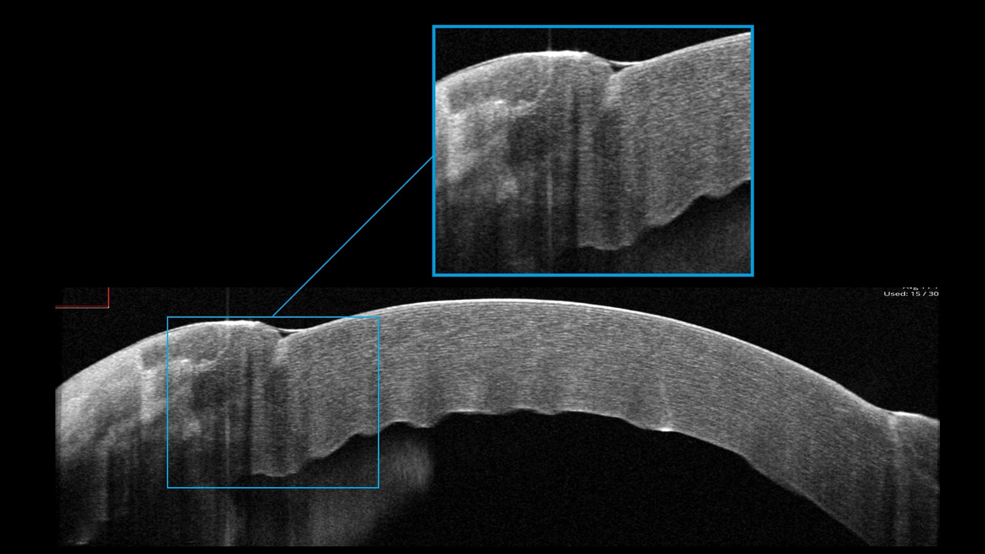
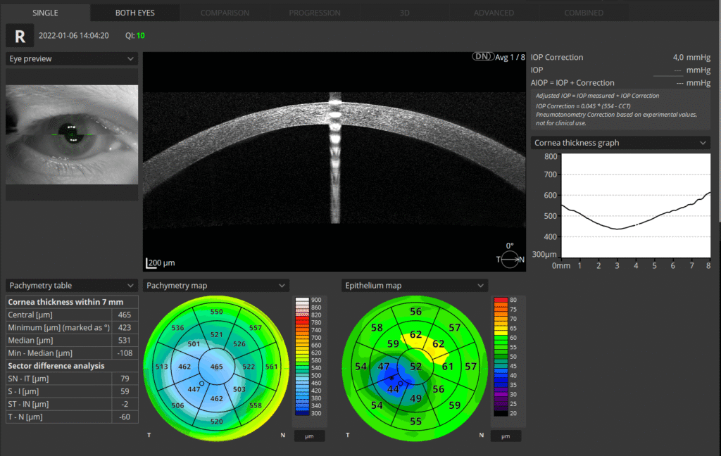
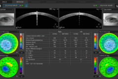
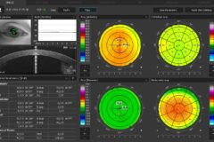
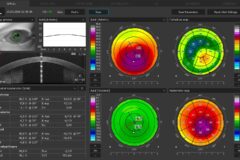
AI Anterior Segmentation
Cornea, Epithelium, Stroma
AI-supported analysis of the anterior segment enables detailed evaluation of corneal structures, including epithelium and pachymetry.
Presentation of results for both eyes supports quick and precise assessment, while epithelial and pachymetry maps are included as part of the standard package.

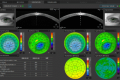
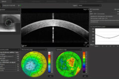
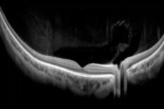
Improved OCT Topography *
OCT Topography provides detailed corneal mapping based on high-resolution OCT data, enabling precise evaluation of corneal thickness distribution and structural profile.
The updated Topography algorithm (T2) enhances corneal surface reconstruction through improved modeling, increasing resistance to noise and improving the reliability of computed parameters. A higher number of B-scans supports more accurate data acquisition and analysis.
T-OCT™ is excellent when paired with the B-OCT® module for IOL surgery.
* - An optional software module

Cataract mode
The cataract mode in the REVO series opens up new possibilities for patients with challening cases.
This featureprovides visualisation of structures hidden beneath opaque layers, making it ideal for diagnosing eye conditions thatwere previously difficult or impossible to study in patients with cataract, corneal oedemas or very dense floaters.The cataract mode allows the scanning speed and sensitivity of the OCT to be modified for better visualisation ofpatients with opaque media.
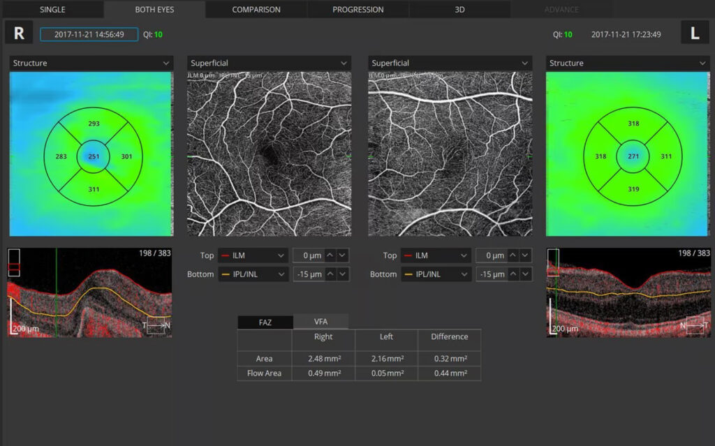
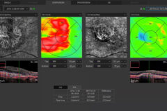
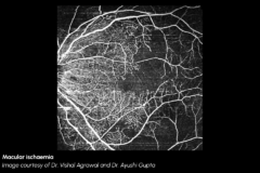
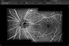
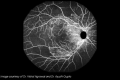
OCT Angiography
This non-invasive dye free technique enables the visualization of the microvasculature of the retina.
OCT Angiography provides an alternative to the traditional fluorescein method. Although OCT-A will not completely replace FA imaging, it is a quick and non-invasive tool. The software allows clinicians to observe, track, and compare changes in the microvasculature of the retina in both eyes.


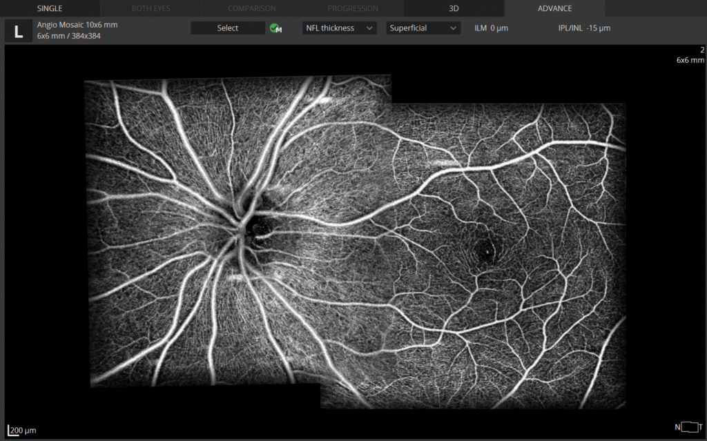
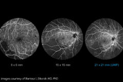
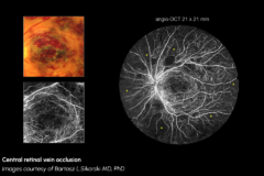
Angiography Mosaic
OCT Angiography Mosaic enables widefield visualization of retinal microvasculature by combining multiple OCT-A scans into a single composite image.
This approach allows clinicians to extend the field of view beyond standard scan sizes — with scan ranges from 3 × 3 mm up to 15 × 15 mm available within a single examination.
It supports a more comprehensive assessment of vascular structures across larger retinal areas.
The mosaic function maintains the advantages of OCT Angiography — non-invasive, dye-free imaging — while enabling broader visualization of microvascular changes.
The software supports visualization, monitoring, and comparison of vascular patterns over time, facilitating follow-up and clinical evaluation.
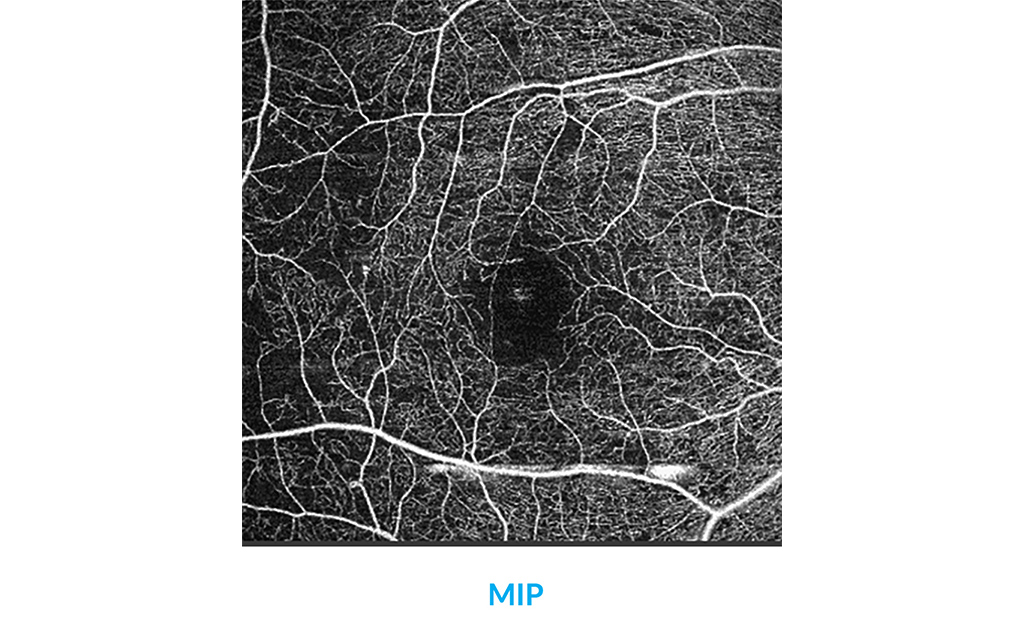
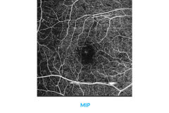
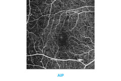
Maximum Intesity Projection
The MIP algorithm
Choose better visualization of angio data for analysis with the Maximum Intensity Projection (MIP) feature. This tool is useful for visualizing OCT-A data as it enables easier identification and tracking of high-intensity structures such as blood vessels.

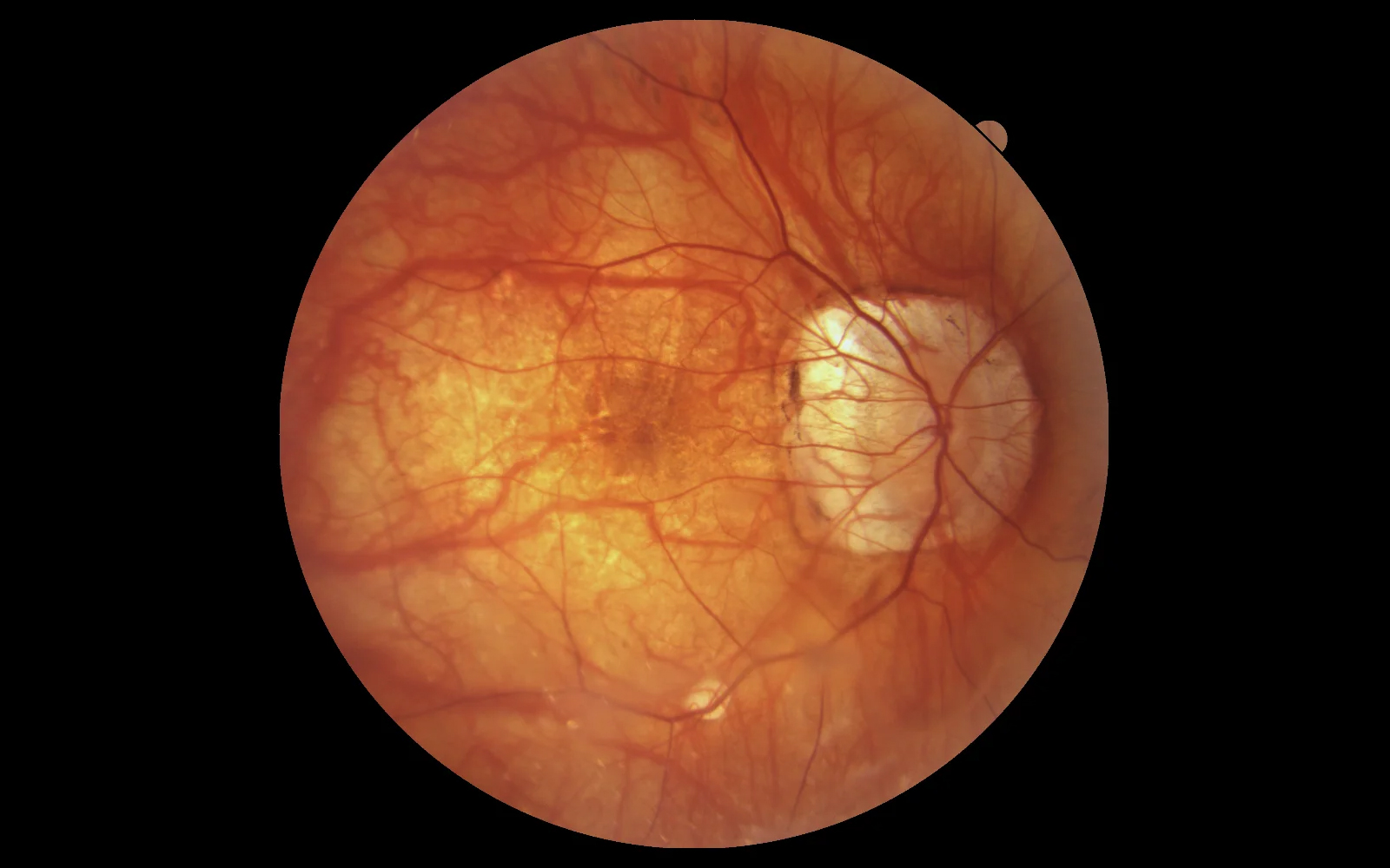
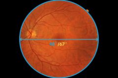
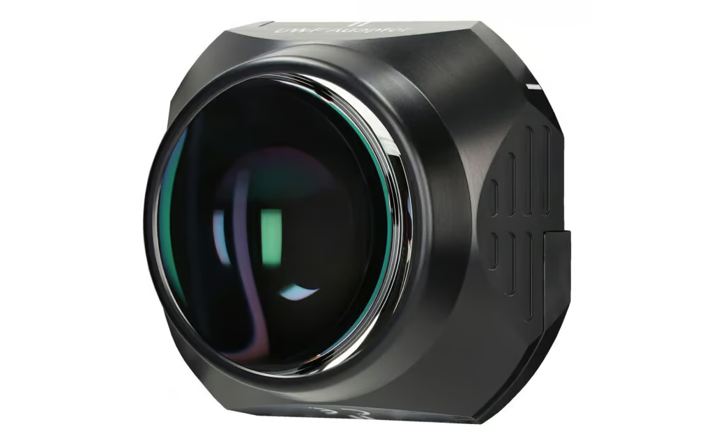
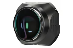
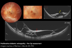
Ultra-Widefield Module
UWF module, available as an optional adapter, provides a new wide perspective of imaging up to approximately 105° with a single scan.
It allows imaging of the macular area along with the far periphery, supporting detection of early-stage pathologies in the posterior segment of the eye.
The module enables 3D imaging for comprehensive analysis, averaging in enhancement mode, and OCT Angiography, allowing visualization of perfusion changes in the peripheral retina.
With the UWF feature in Extended Depth™ software, assessment of high myopia patients becomes more effective when combined with Full Range scanning technology.
For patients with fixation difficulties, the UWF module also provides a Radial scan option, reducing acquisition time and facilitating reliable signal capture.

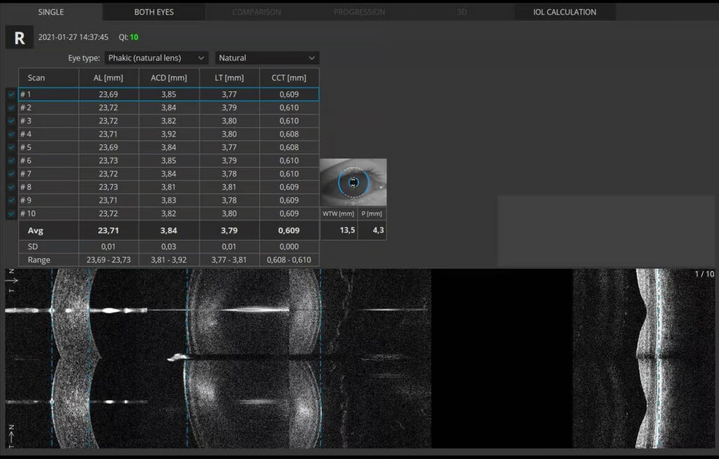
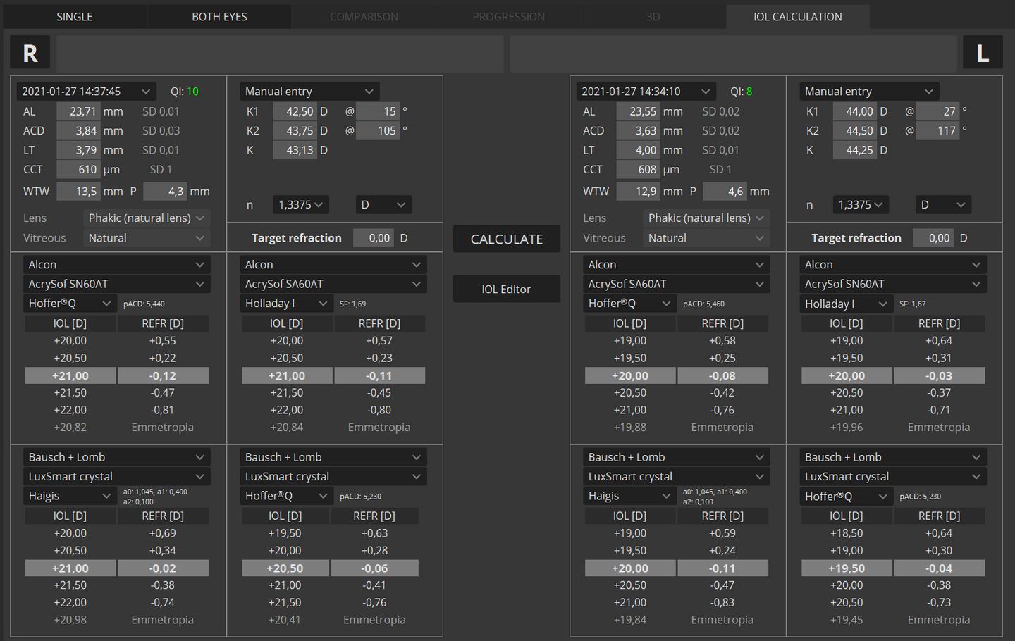
OCT Biometry
B-OCT® is an excellent tool for any clinician who manages myopia control or performs cataract surgery. Biometry OCT provides a complete set of ocular parameters: Axial Length, Central Cornea Thickness, Anterior Chamber Depth, Lens Thickness, Pupil size, and White to White.

High Myopia
The Myopia Forecast module opens progression of the ocular structure parameters according to trends over population mode. Usage reference based on research from multiple universities along with environmental factors allow the monitoring of changes from childhood to adolescence.
The REVO offers an exclusive selection of reference data based on different studies over different time frames and demographics. Reference data can be selected from the NICER San Diez or Tideman studies.


IOL Calculator
OL formulas allow the user to calculate IOL implant parameters. Our systems now support the latest IOL data base standard IOLCon.org so that you can always keep your library up-to-date.
IOL Formulas*:
- Hoffer® Q,
- Holladay I,
- Haigis,
- Theoretical T,
- Regression II.

