OCT Angiography
OCT Angiography
This non-invasive dye free technique enables the visualization of the microvasculature of the retina.
OCT Angiography provides an alternative to the traditional fluorescein method. Although OCT-A will not completely replace FA imaging, it is a quick and non-invasive tool. The software allows clinicians to observe, track, and compare changes in the microvasculature of the retina in both eyes.
OCT-A Analysis Methods
Quantification
The quantification tool provides quantification of the vasculature in the whole analyzed area together with values in specific zones and sectors. Using the heat map of the analyzed vasculature, the evaluation of vascular structure conditions is much faster. The choice of the quantification method increases the sensitivity of analyses for specific deseases.
Available quantification methods:
- Vessel Area Density – it is defined as the total area of perfused vasculature per unit area in a region of measurement.
- Skeleton Area Density – it is defined as the total area of skeletonized vasculature per unit area in a region of measurement.
Quantification is available for a specific layer in Angio OCT exam:
- Retina: Superficial Plexus and Deep Plexus
- Disc: RPC – Radial Peripapillary Capillary
OCT-A Analytical Tools
OCT-A Mosaic
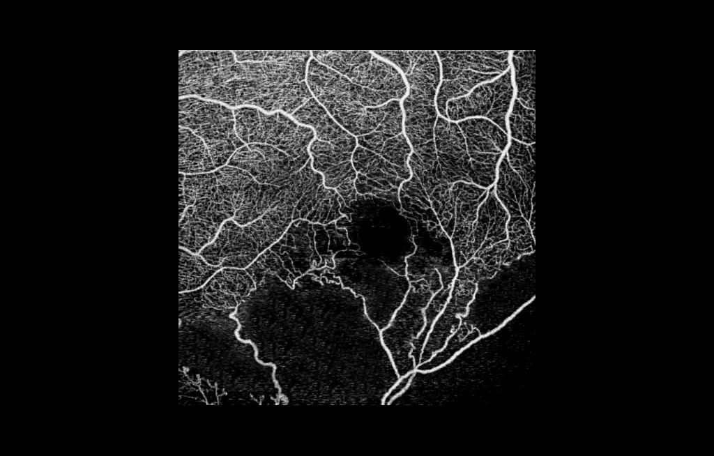
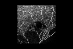
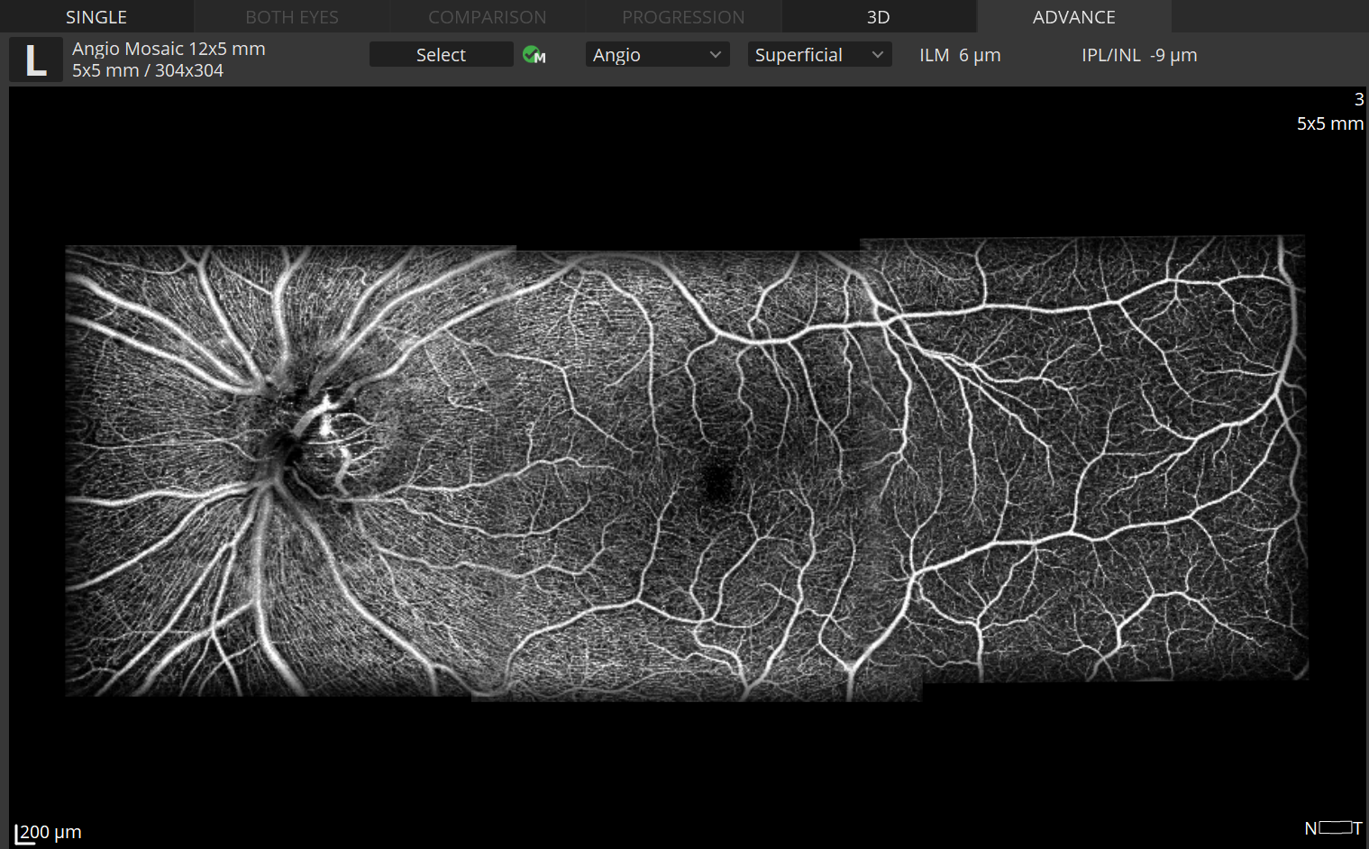
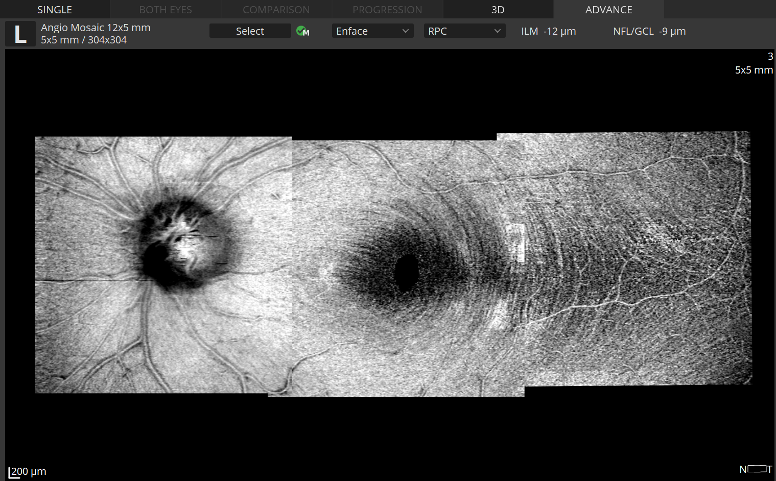
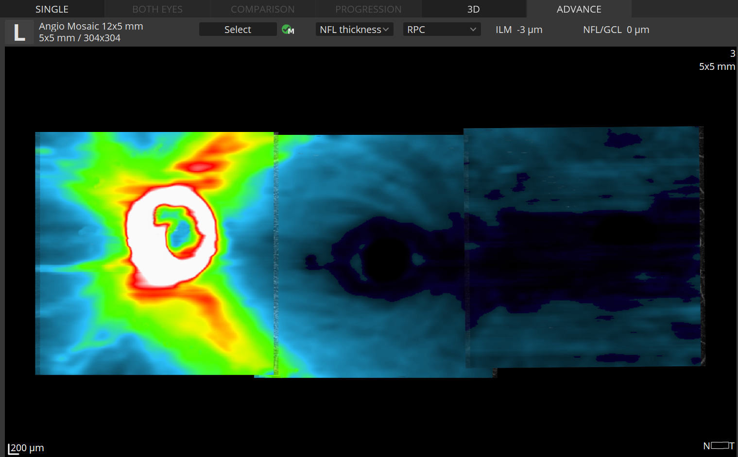
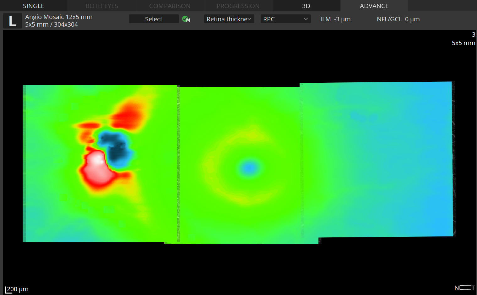
The Angiography mosaic delivers high-detailed images over large field of the retina.
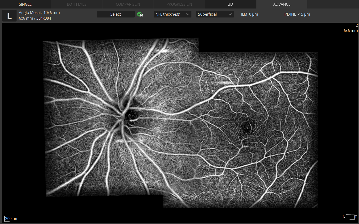
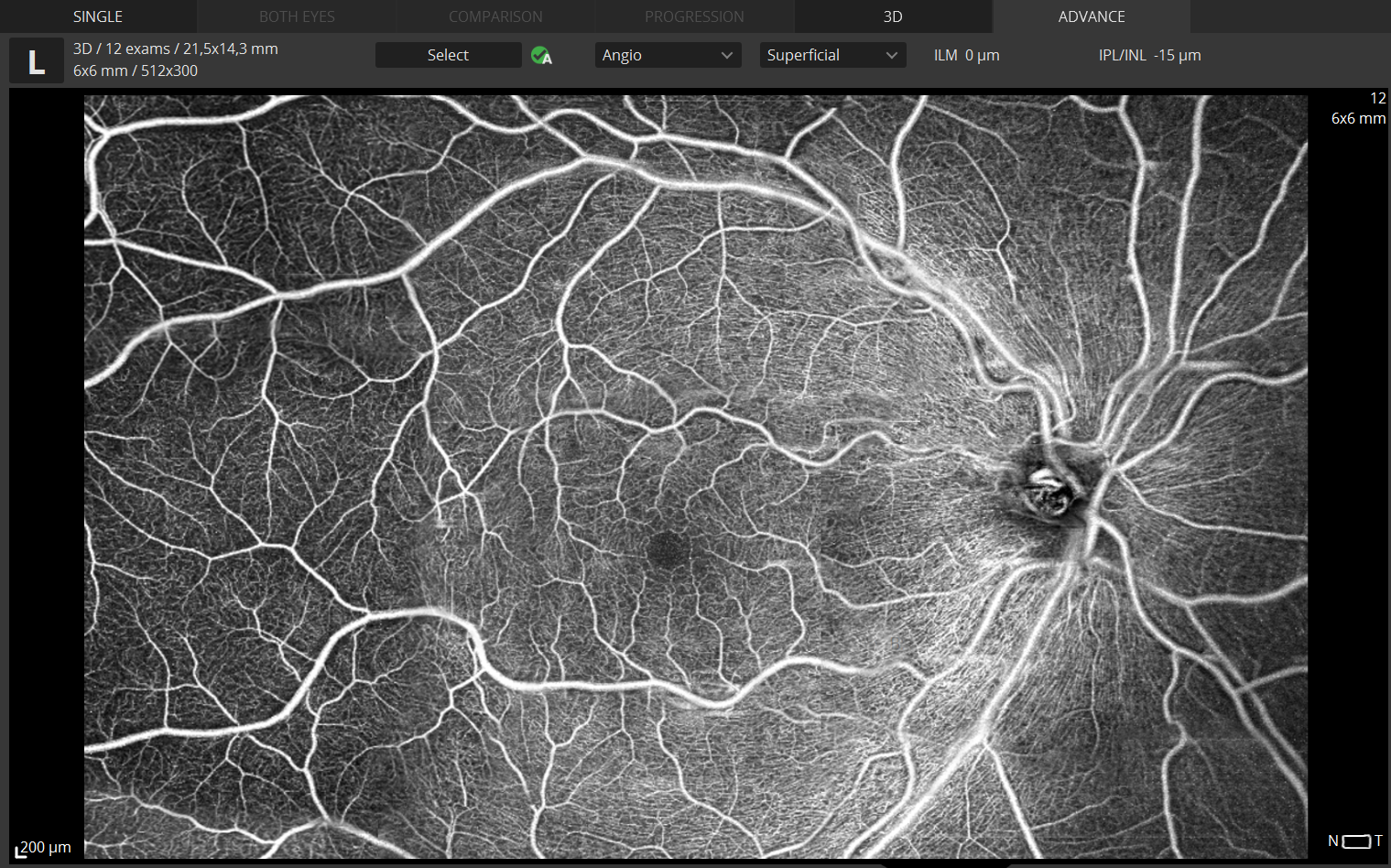
Advanced tab:
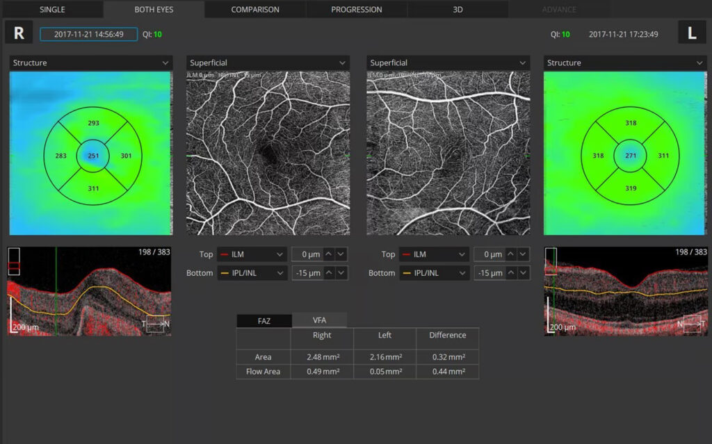
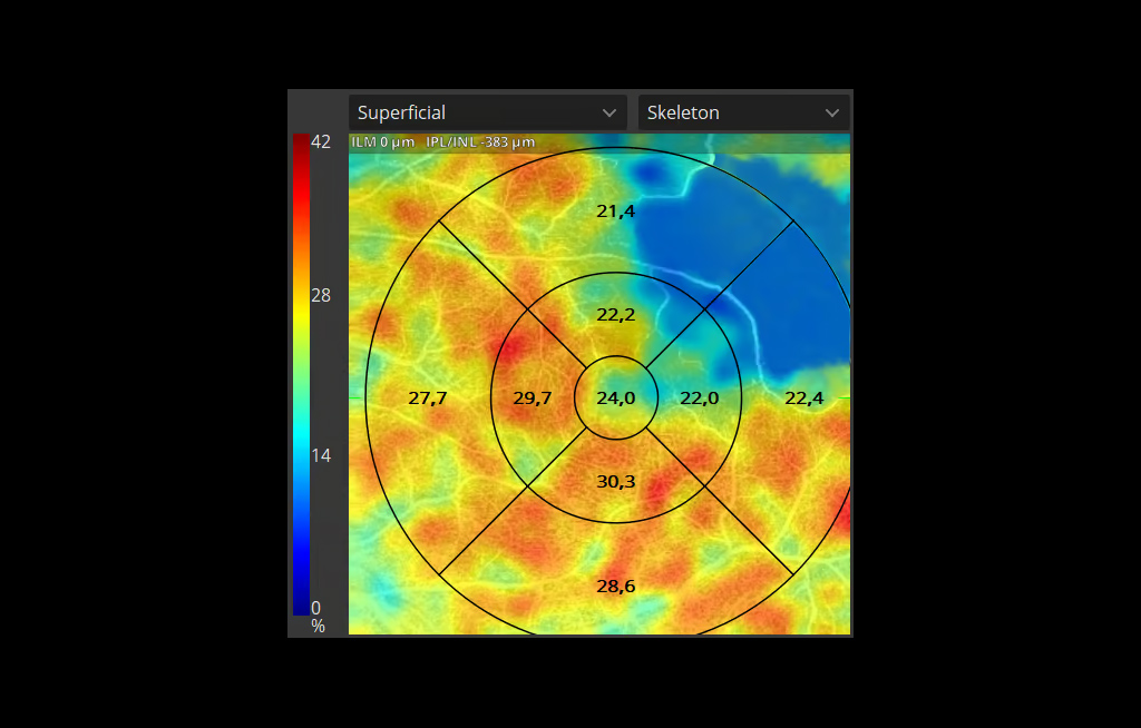
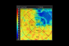
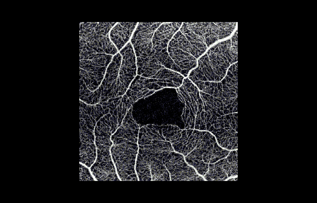
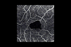
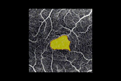
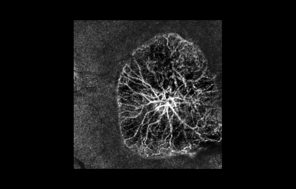
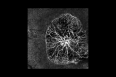
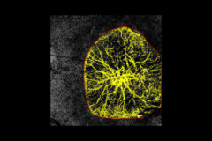
provides view of any vascular layers, enface view of vascular layers, depth coded and thickness map.
Mosaic modes:
10×6 mm, 12×5 mm, 7×7 mm, 10×10 mm and Manual (up to 12 images).
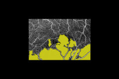
Manual mode:
allows to scan the desired region.Built-in analytics allow to see vascular layers, enface or thick-ness maps.