| OPTICAL COHERENCE TOMOGRAPHY |
| Technology |
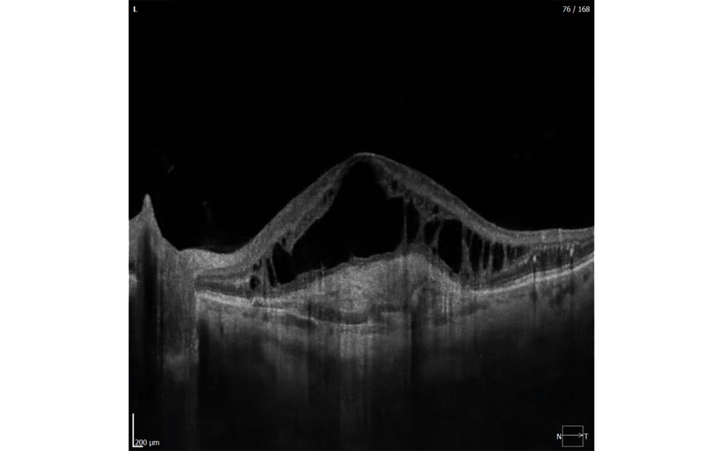
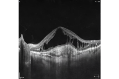
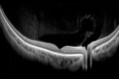
Spectral Domain OCT |
| Light source |
SLED 870 nm, 93 nm width |
| Bandwidth |
93 nm half bandwidth |
| Scanning speed |
130 000 A-scan/sec |
| Axial resolution |
~1.3 μm digital, 3 μm in tissue |
| Transverse resolution |
12 μm, typically 18 μm |
| Overall scan depth |
2.6 mm / 5.4 mm in Full Range mode |
| Min. pupil size for OCT |
1.7 mm |
| Focus adjustment range |
-25 D to +25 D |
| Scan range |
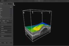
Posterior 3–15 mm, Angio 3–15 mm, Anterior 3–18 mm |
| Scan types |
3D, Angio², Full Range Radial, Full Range B-scan, Radial (HD), B-scan (HD), Raster (HD), Raster 21 (HD), Cross (HD), TOPO², Biometry AL² |
| Fundus alignment |
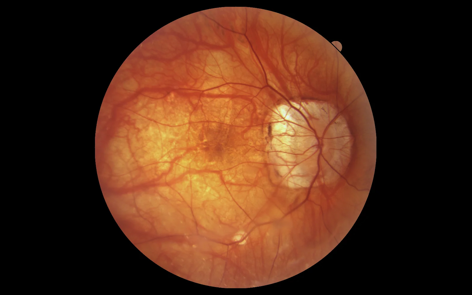
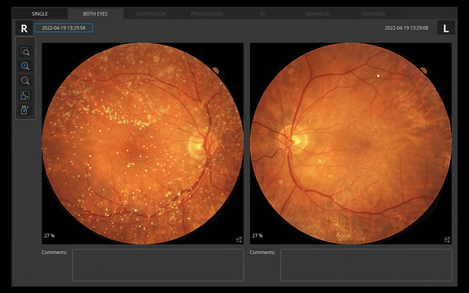
IR, pSLO (Live Fundus Reconstruction) |
| Alignment method |
Fully automatic, Automatic, Manual |
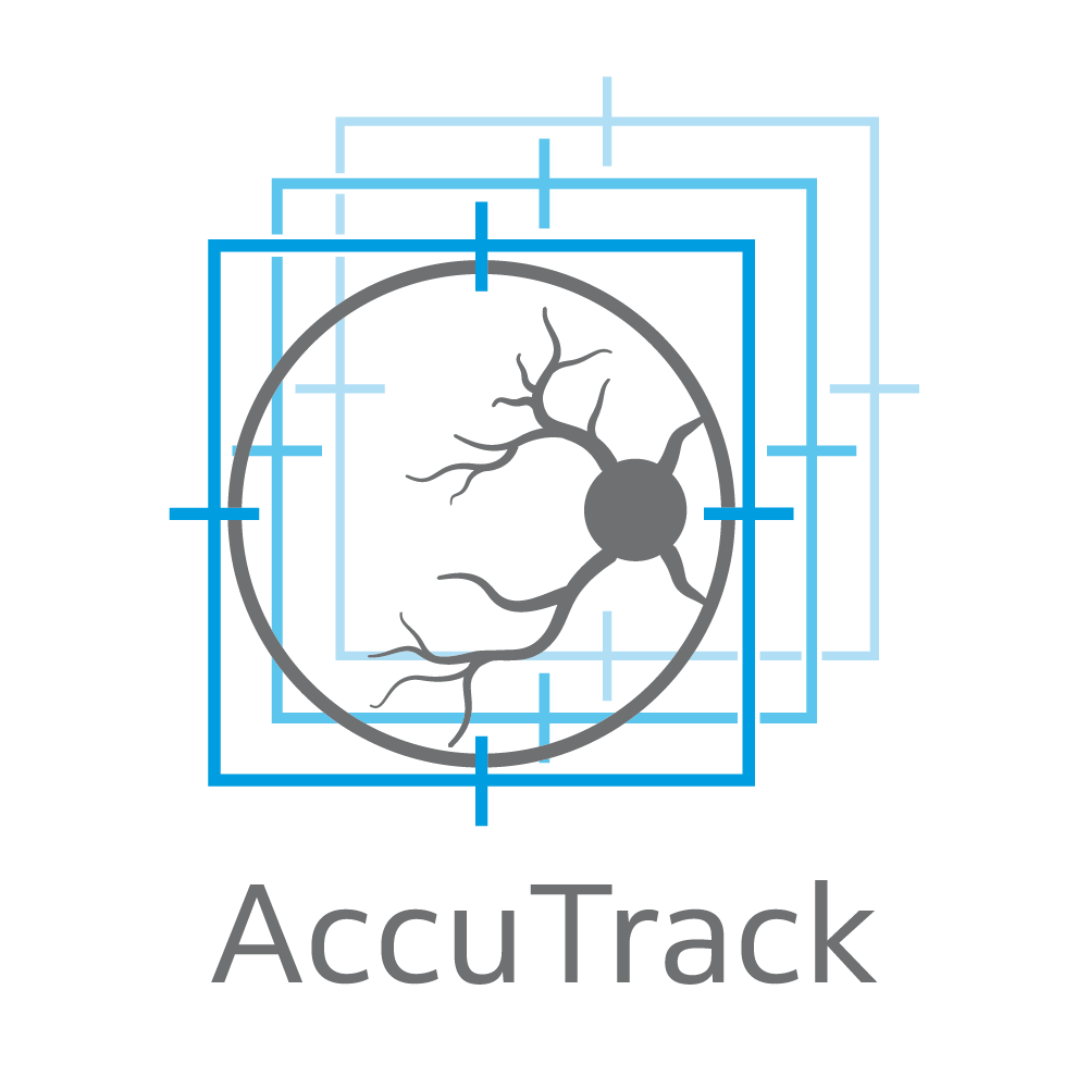
| Fundus tracking |
ACCUtrack – active real time, iTracking |
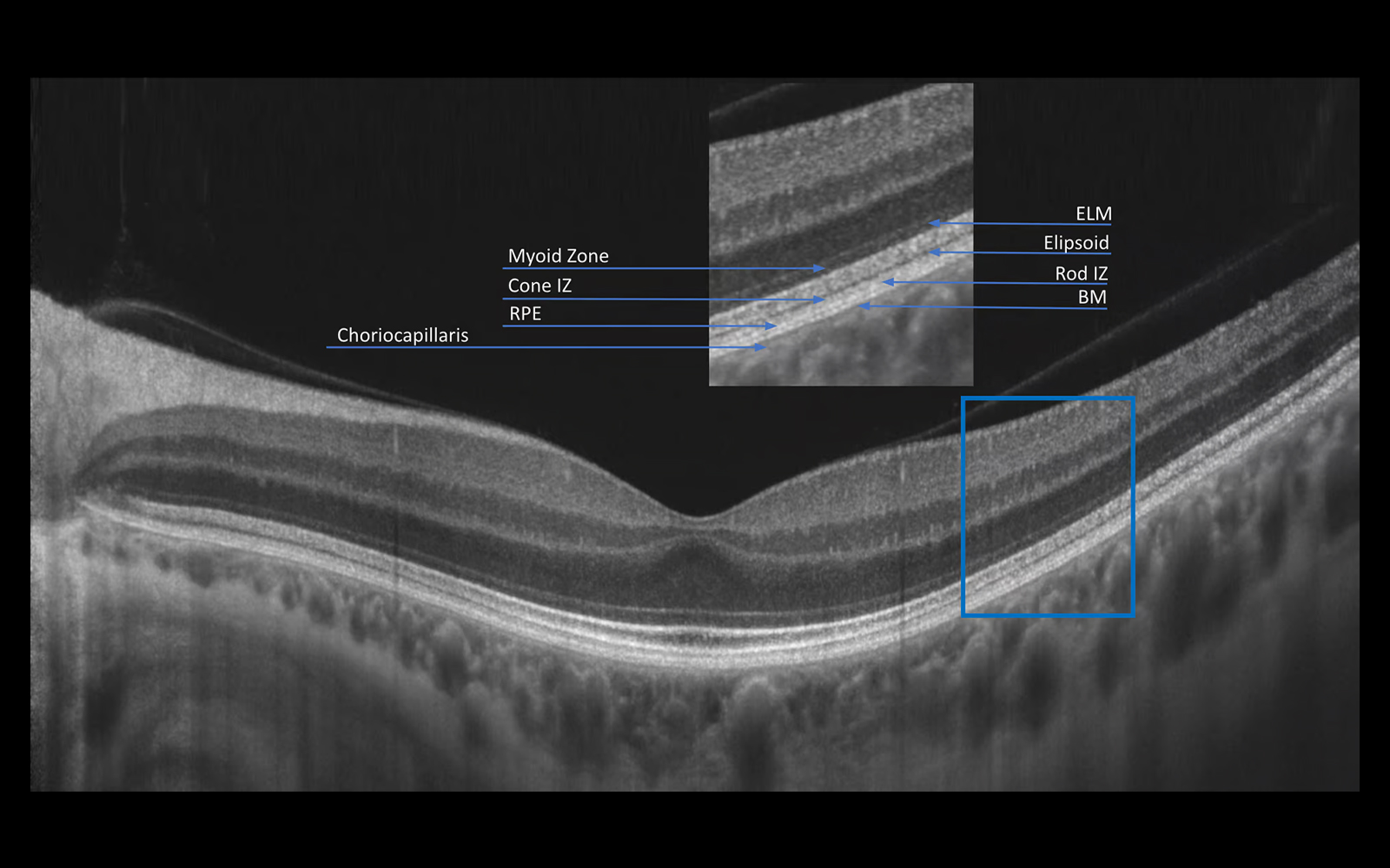
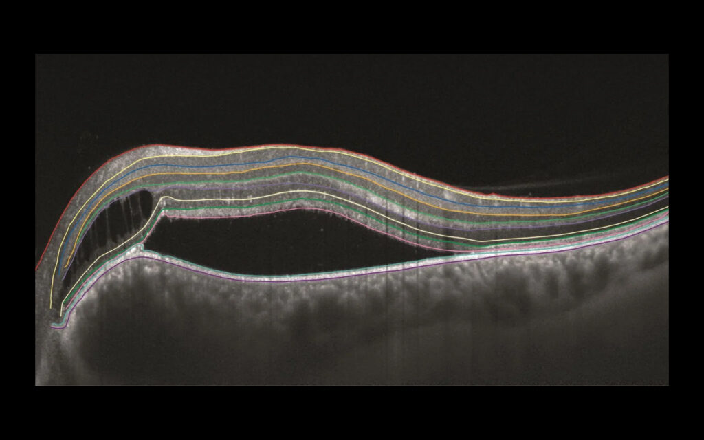
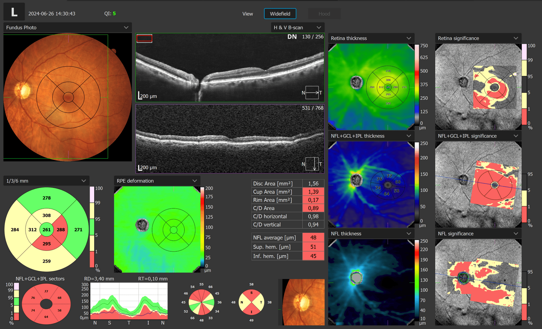
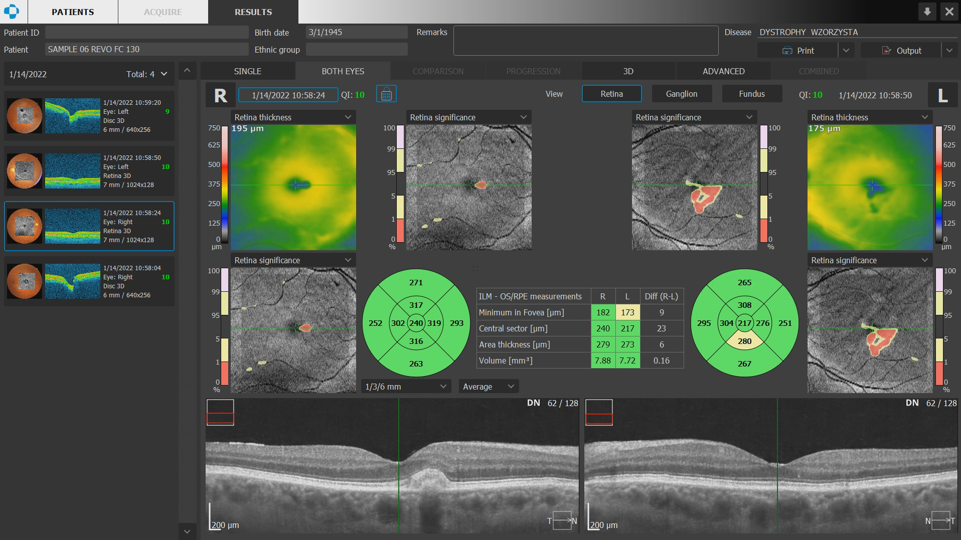
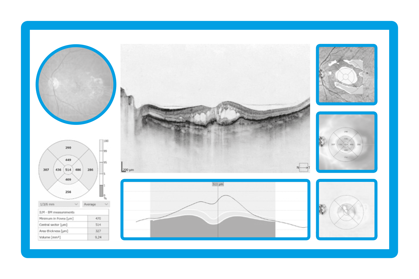
| Retina analysis |
Retina thickness, Inner Retinal thickness, Outer Retinal thickness, RNFL+GCL+IPL thickness, GCL+IPL thickness, RNFL thickness, RPE deformation, MZ/EZ-RPE thickness |
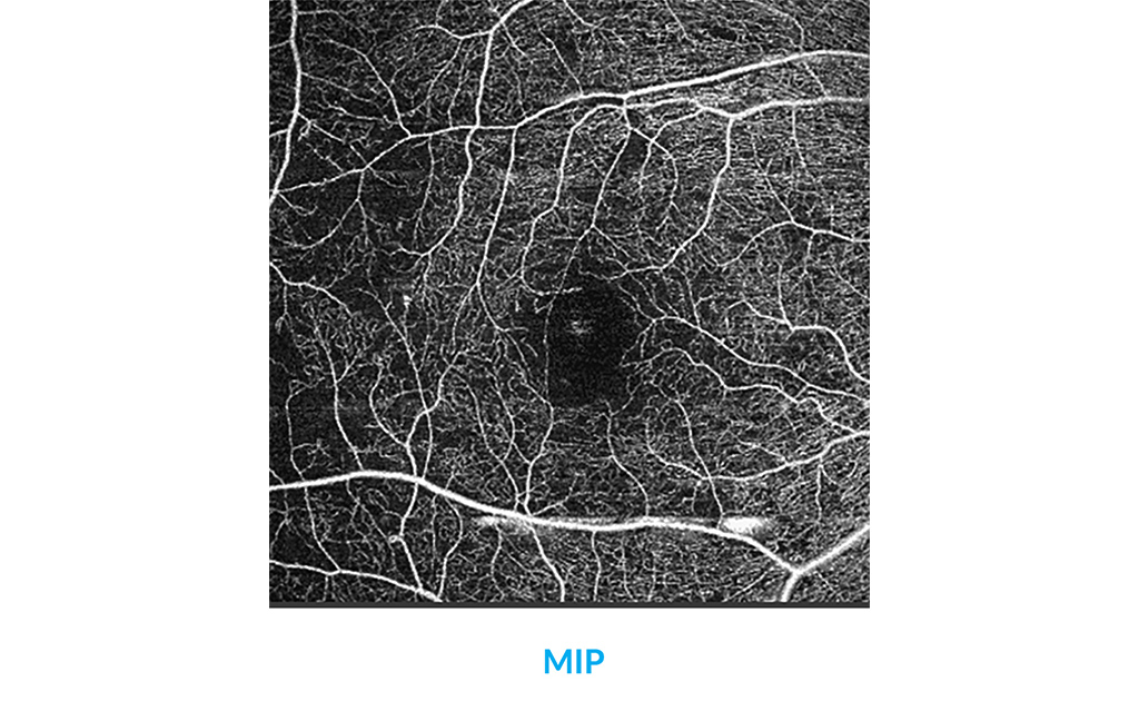
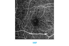
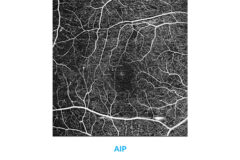
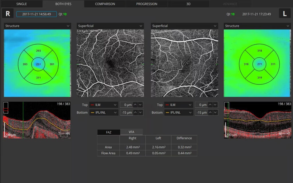
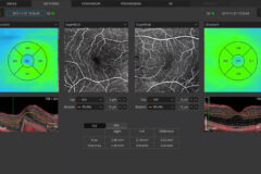
| Angiography OCT² |
Vitreous, Retina, Choroid, Superficial Plexus, RPCP, Deep Plexus, Outer Retina, Choriocapilaries, Depth Coded, SVC, DVC, ICP, DCP, Custom, Enface
Quantification: FAZ, VFA, NFA, Vessel Area Density, Skeleton Area Density, Thickness map |
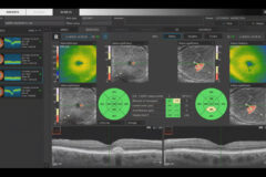
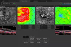
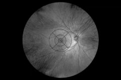
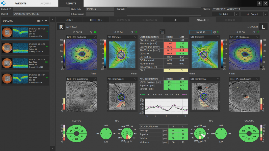
| Glaucoma analysis |
RNFL, ONH morphology, DDLS, OU and Hemisphere asymmetry, Ganglion analysis as RNFL+GCL+IP and GCL+IPL, Structure + Function¹ |
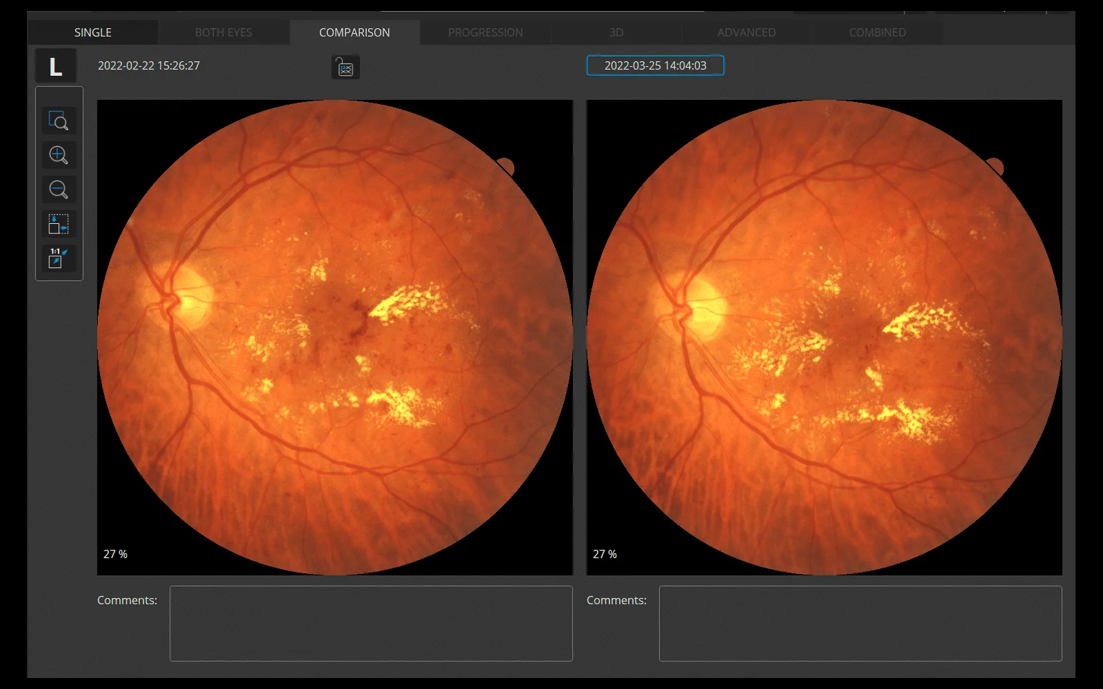
| Angiography mosaic |
Acquisition: Auto, Manual; Mosaic modes: 10×10, 10×6, 12×5, 7×7, Manual up to 12 images |
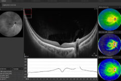
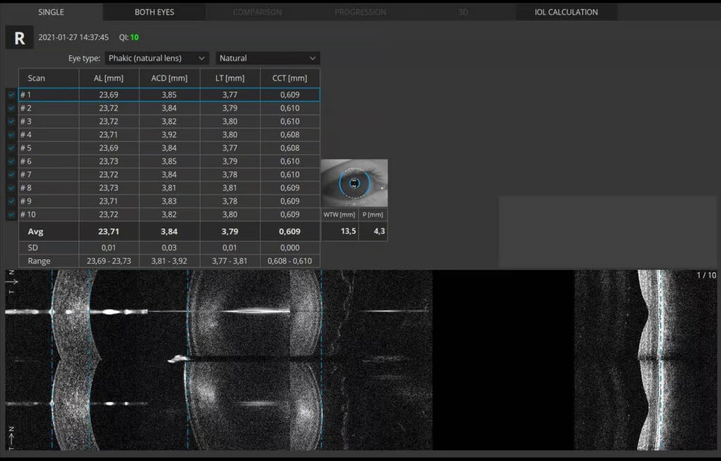
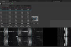
| Biometry OCT² |
AL, CCT, ACD, LT, P, WTW |
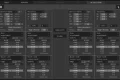
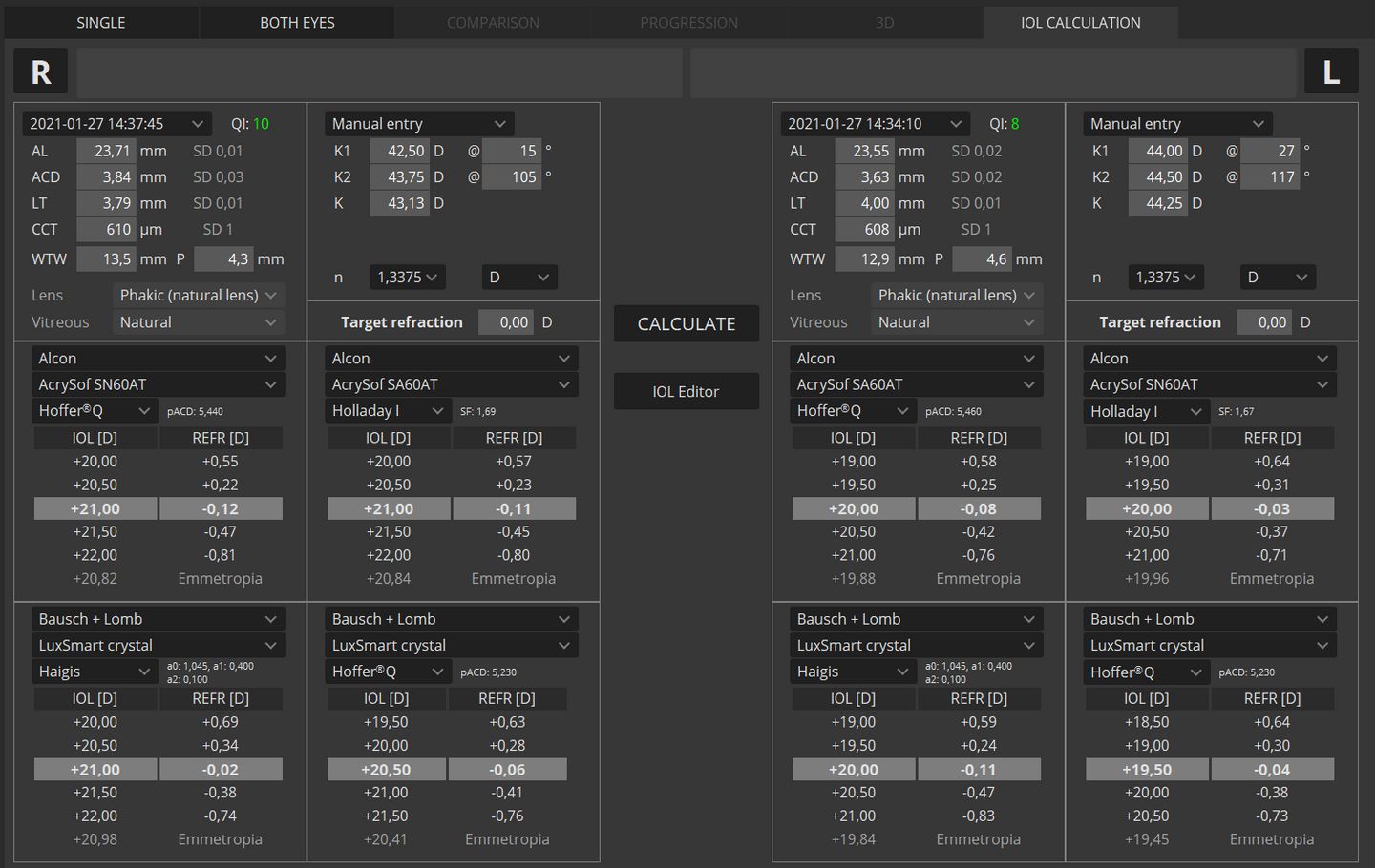
| IOL Calculator³ |
Hoffer Q, Holladay I, Haigis, Theoretical T, Regression II |
| Corneal Topography Map² |
Axial [Anterior, Posterior], Refractive Power [Kerato, Anterior, Posterior, Total], Net Map, Axial True Net, Equivalent Keratometer, Elevation [Anterior, Posterior], Height, KPI (Keratoconus Prediction Index) |
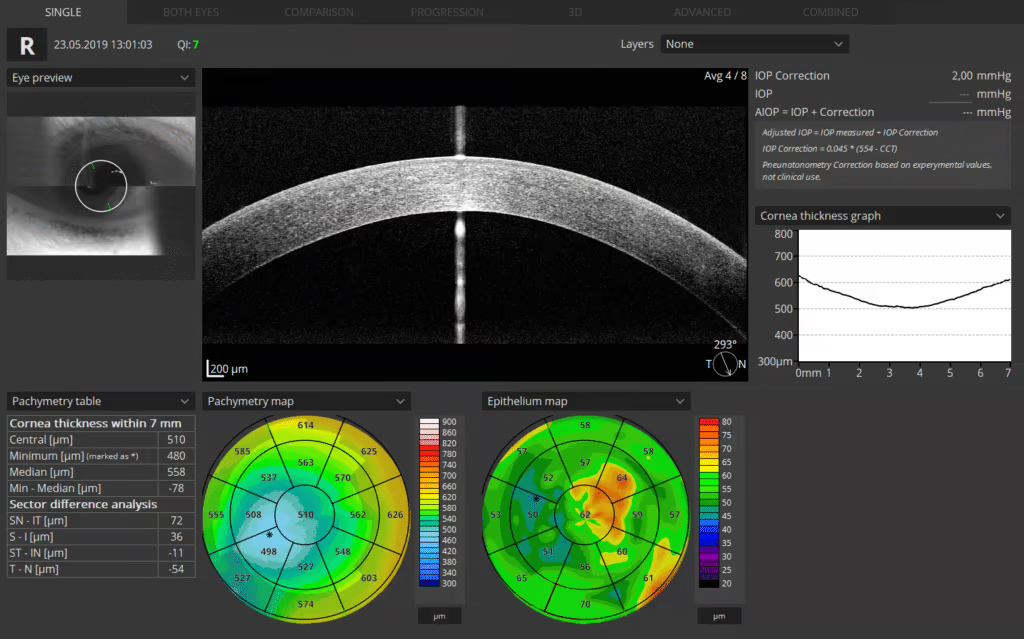
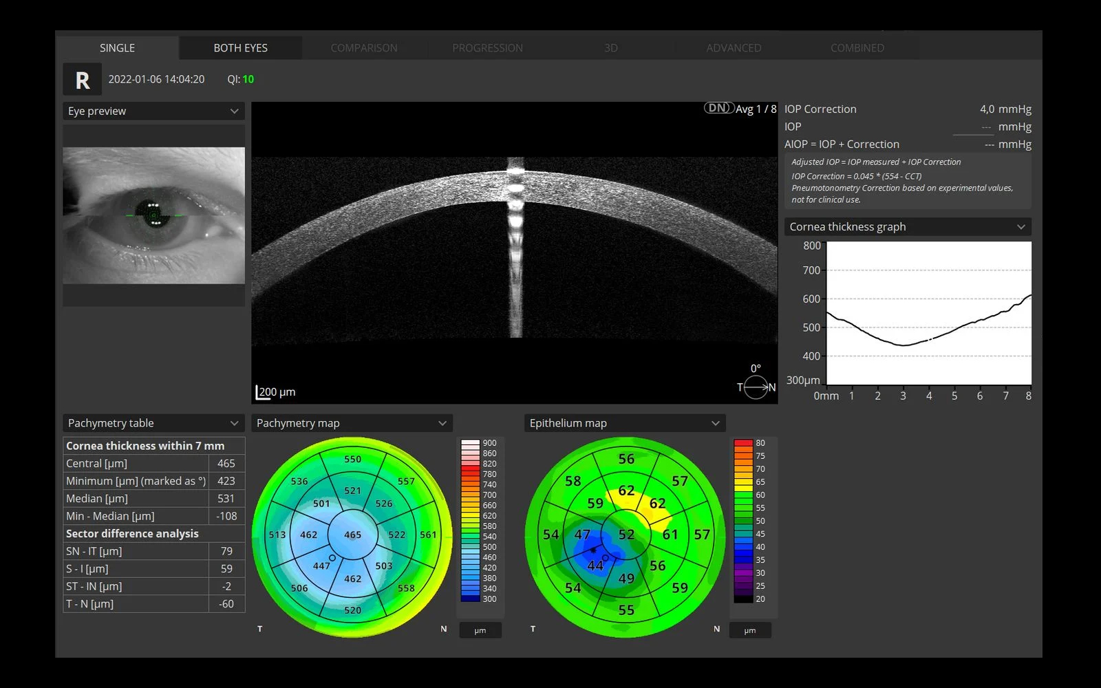
| Anterior (no lens/adapter required) |
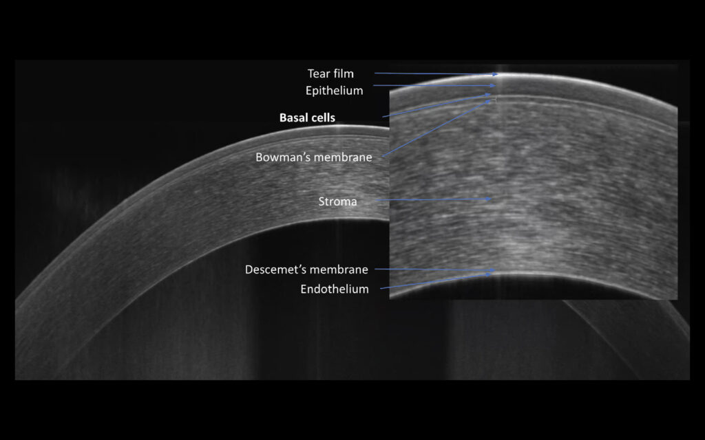
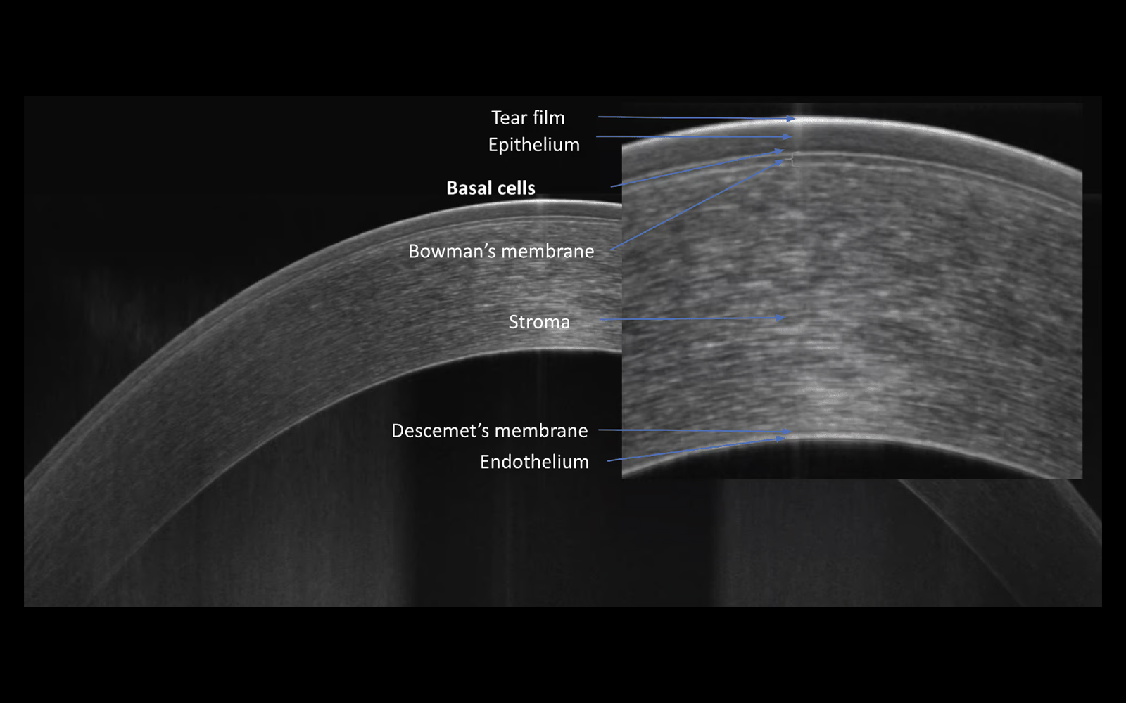
Anterior Chamber Radial, Anterior Chamber B-scan, Pachymetry, Epithelium map, Stroma map, Angle Assessment, AIOP, AOD 500/750, TISA 500/750, Angle to Angle view |
| Connectivity |
DICOM Storage SCU, DICOM MWL SCU, CMDL, Networking |
| Fixation target |
OLED display (the target shape and position can be changed), External fixation arm |
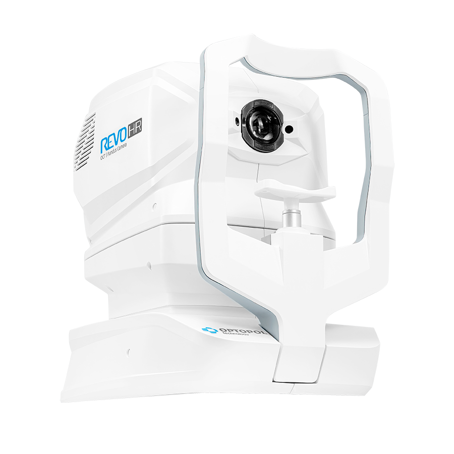
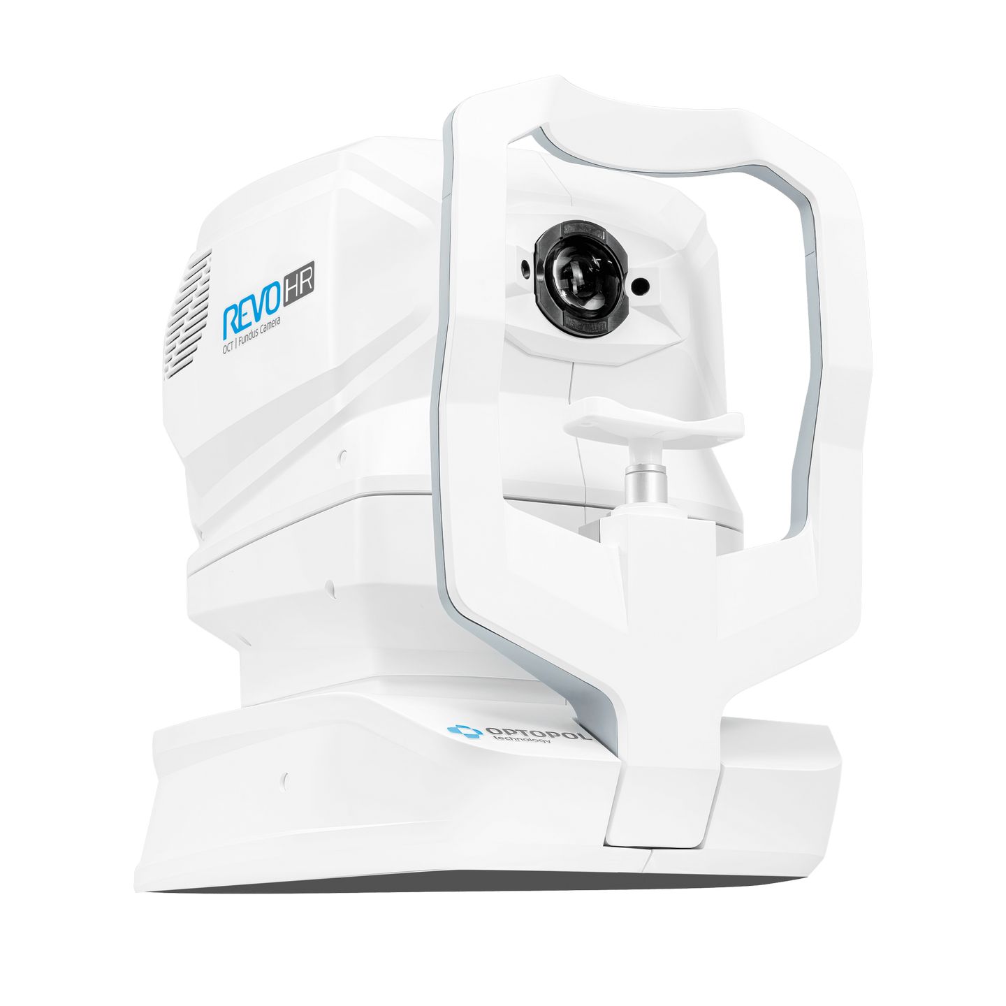
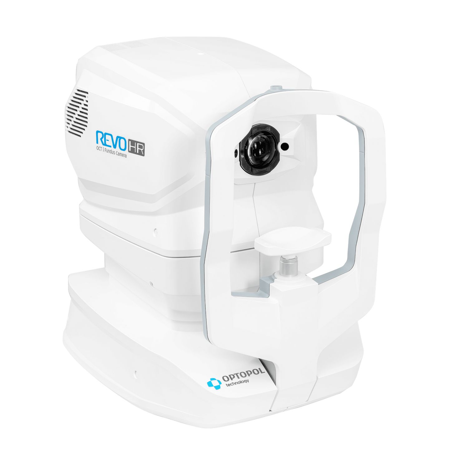
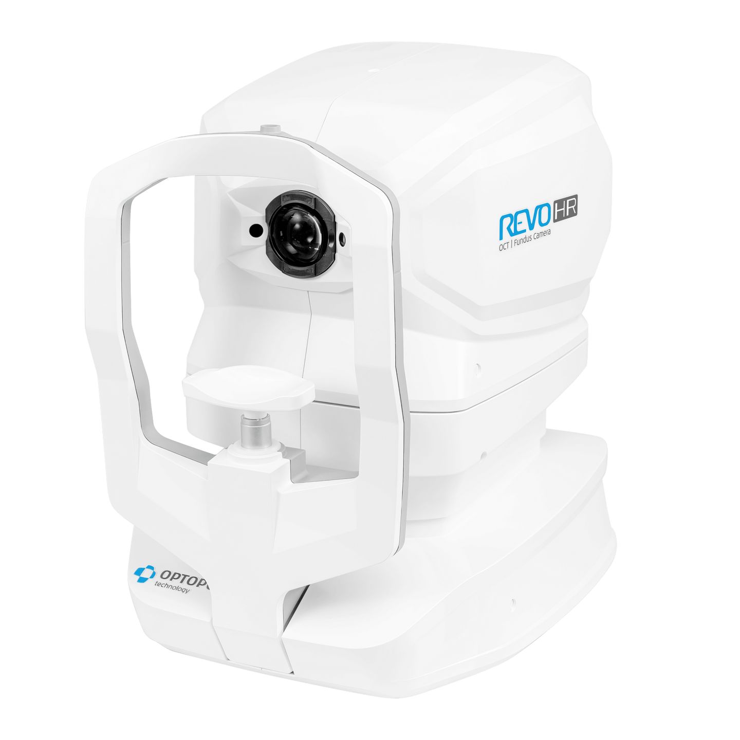
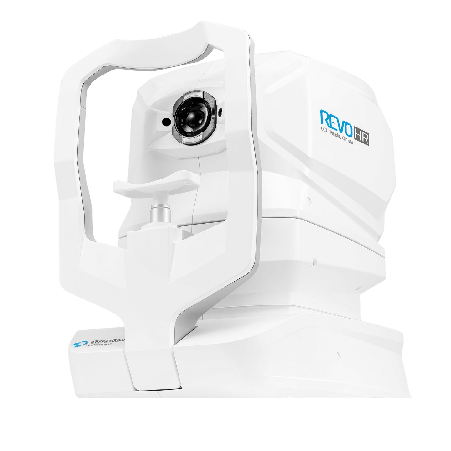
| Dimensions (L×W×H) / Weight |
479 × 367 × 493 mm / 30 kg |
| Power supply / Consumption |
100–240 V, 50/60 Hz / 90–110 VA |


