
COMING SOON
FUNDO
One platform. Every retinal perspective.
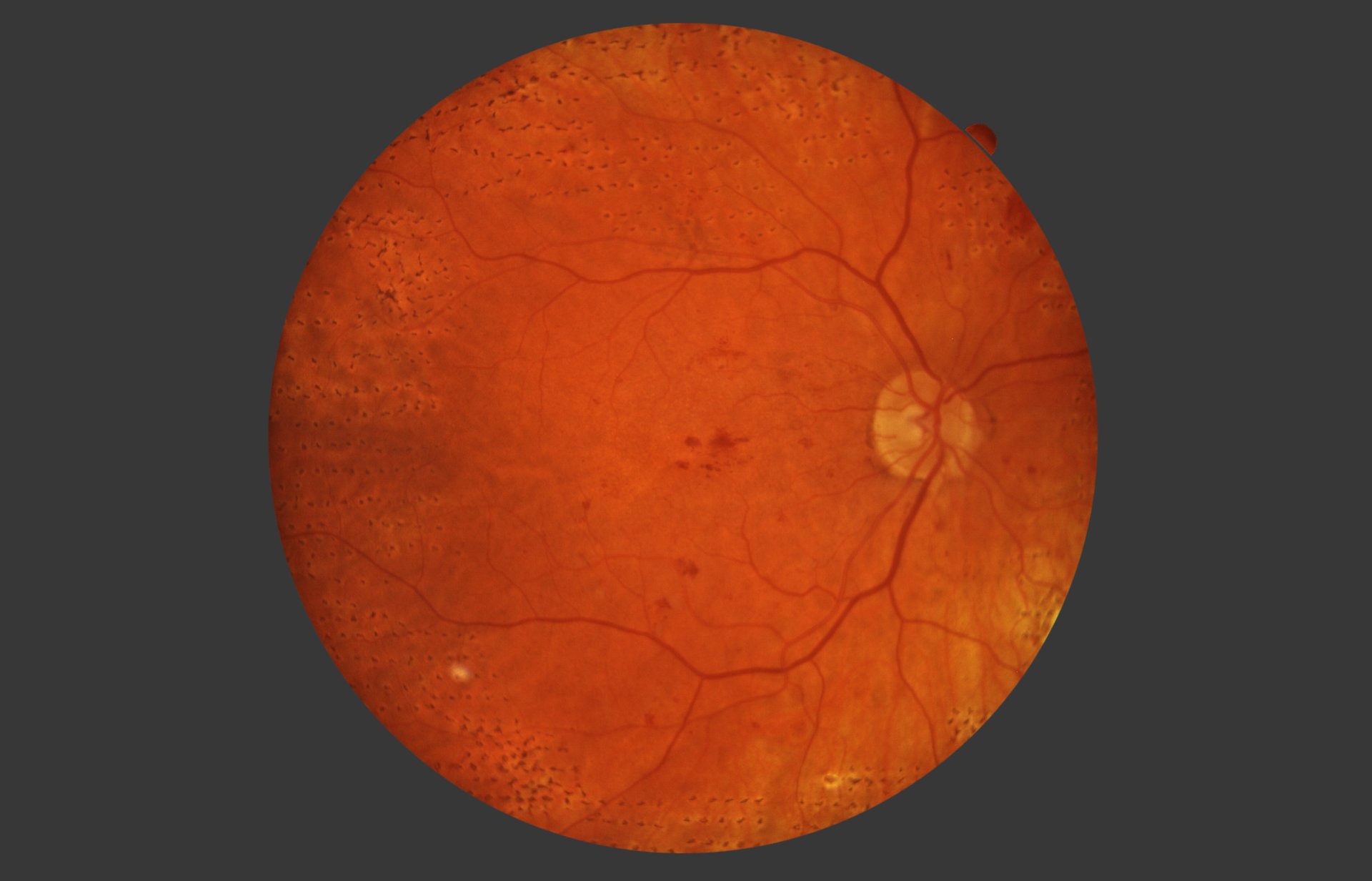
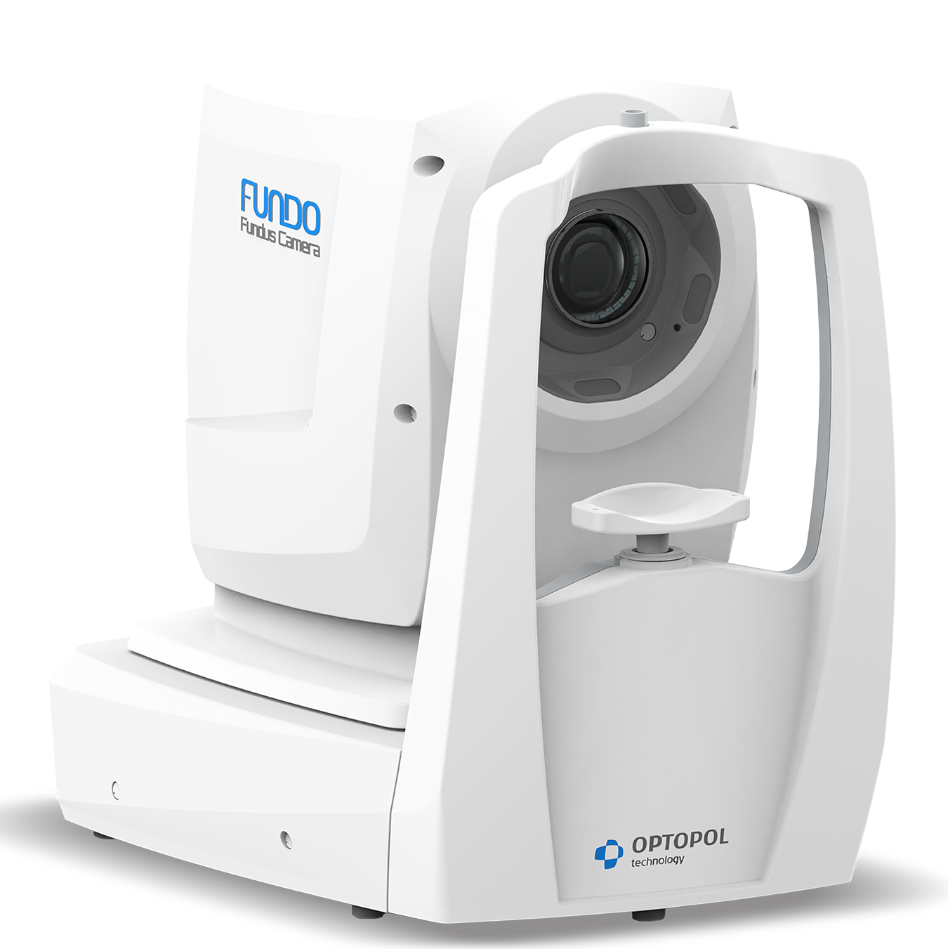
The FUNDO is an automatic, non-mydriatic widefield fundus camera designed for efficient retinal diagnostics. It redefines multimodal fundus and anterior segment imaging by combining widefield retinal imaging, true-color reproduction, fully automated examinations, AI-powered support, and seamless integration into the clinical workflow.
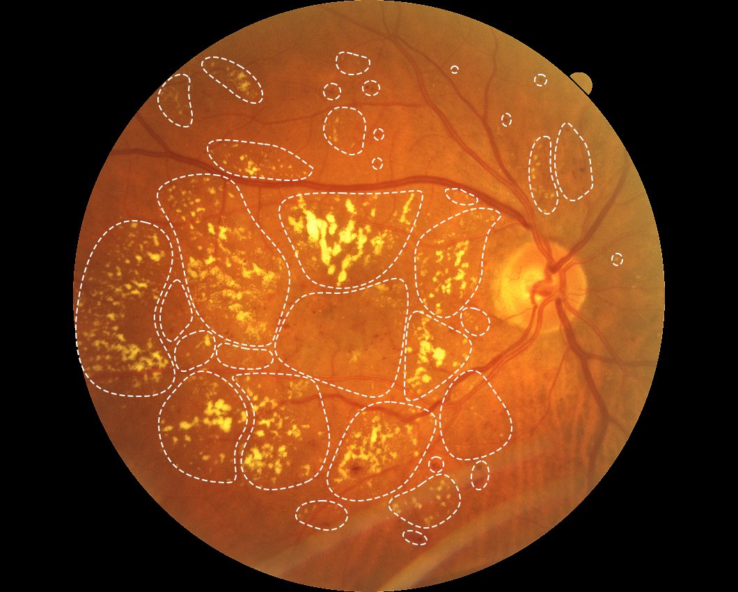
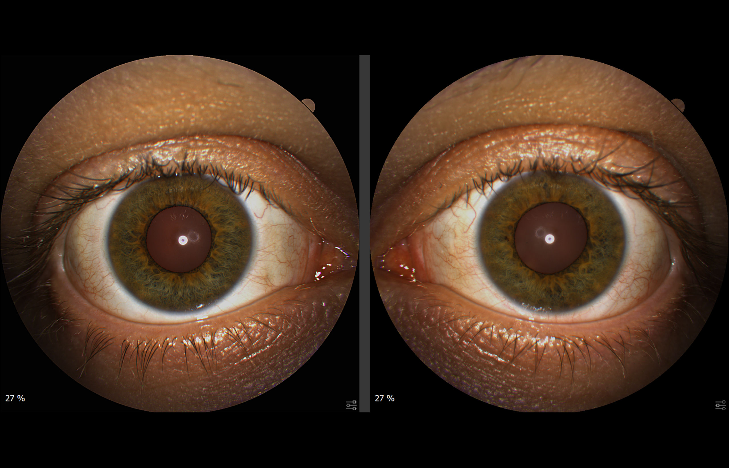
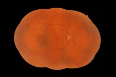
With a single 60° widefield acquisition, the FUNDO captures a broad view of the fundus — from the macula to the retinal periphery. The system provides multimodal imaging, including color fundus photography, infrared fundus imaging, green and blue fundus autofluorescence, fluorescein angiography, indocyanine green angiography, anterior segment color photography, and meibography, supporting detailed assessment of ocular structures across a wide range of clinical applications
Advanced Fundus Imaging For Everyday Clinical Practice
A Wide Perspective Of The Retina
Automation That Saves Time
Comfort Without Routine Mydriasis
Detailed Clinical Imaging
Connected Diagnostics and Flexible Data Export
Ready For Every Practice
Advanced Fundus Analysis Tools
A comprehensive set of image processing and analysis tools enables efficient assessment, documentation, and follow-up of retinal conditions.
Available functions include:
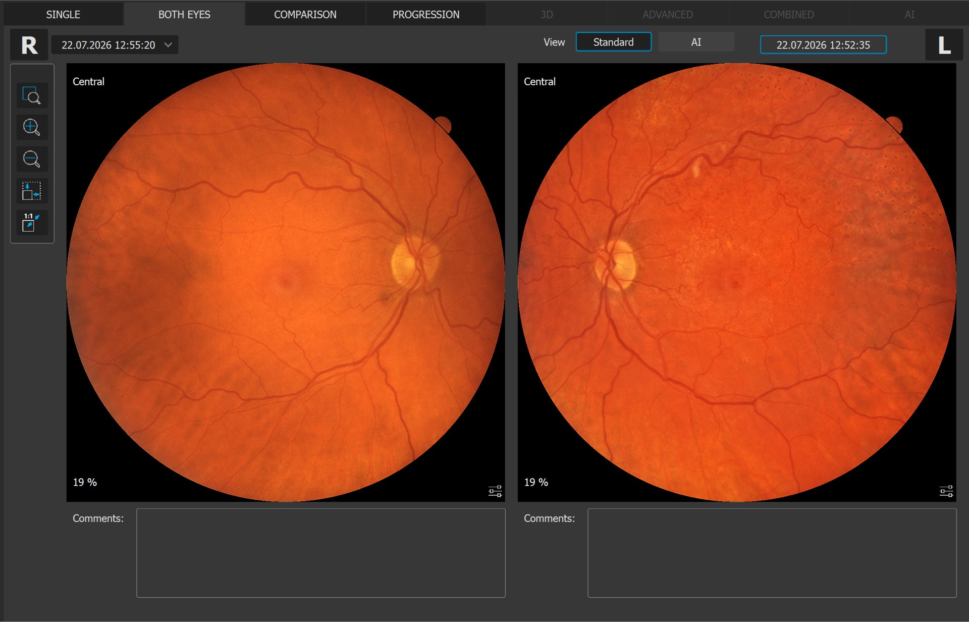
- single-eye and bilateral imaging,
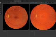
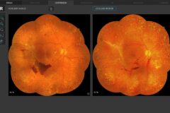
- chronological comparison of fundus photographs, including x4 and x8 progression views, for monitoring disease progression,
- linking a fundus image to multiple REVO and REVO 2 OCT scans and visual field test results (FUNDO, REVO & REVO 2 work on a common patient database).
The set of measurement tools includes Area, Distance, and C/D, while the additional image enhancement filters include Emboss+, Emboss−, Cobalt, Digital Cobalt, and Digital Red-Free.
Multimodal Fundus Imaging
Choose the autofluorescence mode you need – or use both for a more complete assessment.
FAF Green – Green Fundus Autofluorescence
Green fundus autofluorescence supports the assessment of metabolic and structural changes in the retinal pigment epithelium (RPE) and outer retina, particularly in chorioretinal and degenerative retinal disorders.
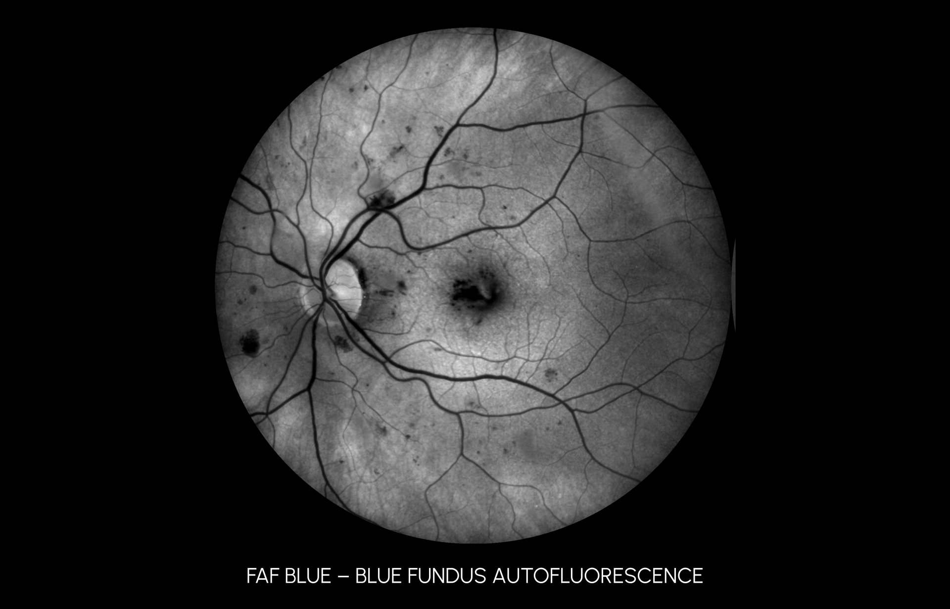
FAF Blue – Blue Fundus Autofluorescence
Blue fundus autofluorescence provides complementary information on RPE integrity, lipofuscin-related changes, and outer retinal abnormalities.
Two angiography modes for deeper vascular insight – retinal with FA, choroidal with ICGA.
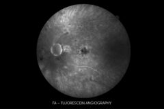
FA – Fluorescein Angiography
Fluorescein angiography enables the assessment of retinal vascular perfusion, leakage, microaneurysms, neovascularization, inflammatory vascular changes, and areas of capillary non-perfusion or ischemia.
ICGA — Indocyanine Green Angiography
Indocyanine green angiography supports the evaluation of choroidal circulation and vasculature, including polypoidal lesions, occult choroidal neovascularization, and deeper chorioretinal vascular abnormalities.
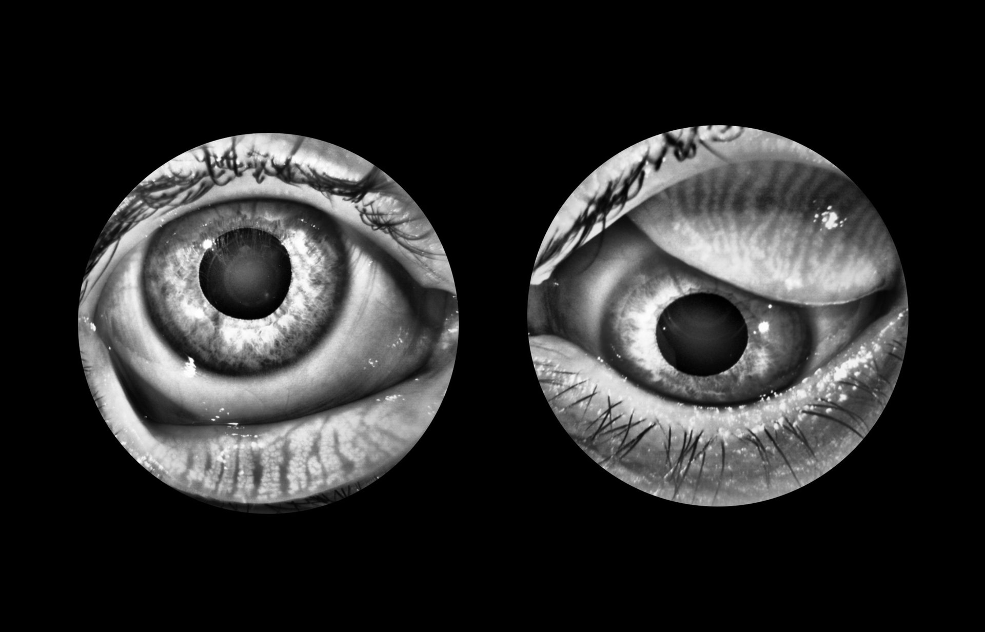
Meibography
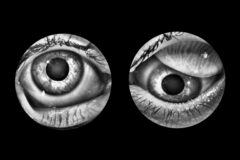
Non-invasive infrared imaging of the Meibomian glands, supporting assessment of their structure and condition. This function helps in the diagnosis of Meibomian Gland Dysfunction (MGD), one of the leading causes of evaporative dry eye disease, and facilitates communication of examination results to the patient.
Ultra-Widefield Fundus Imaging
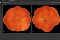
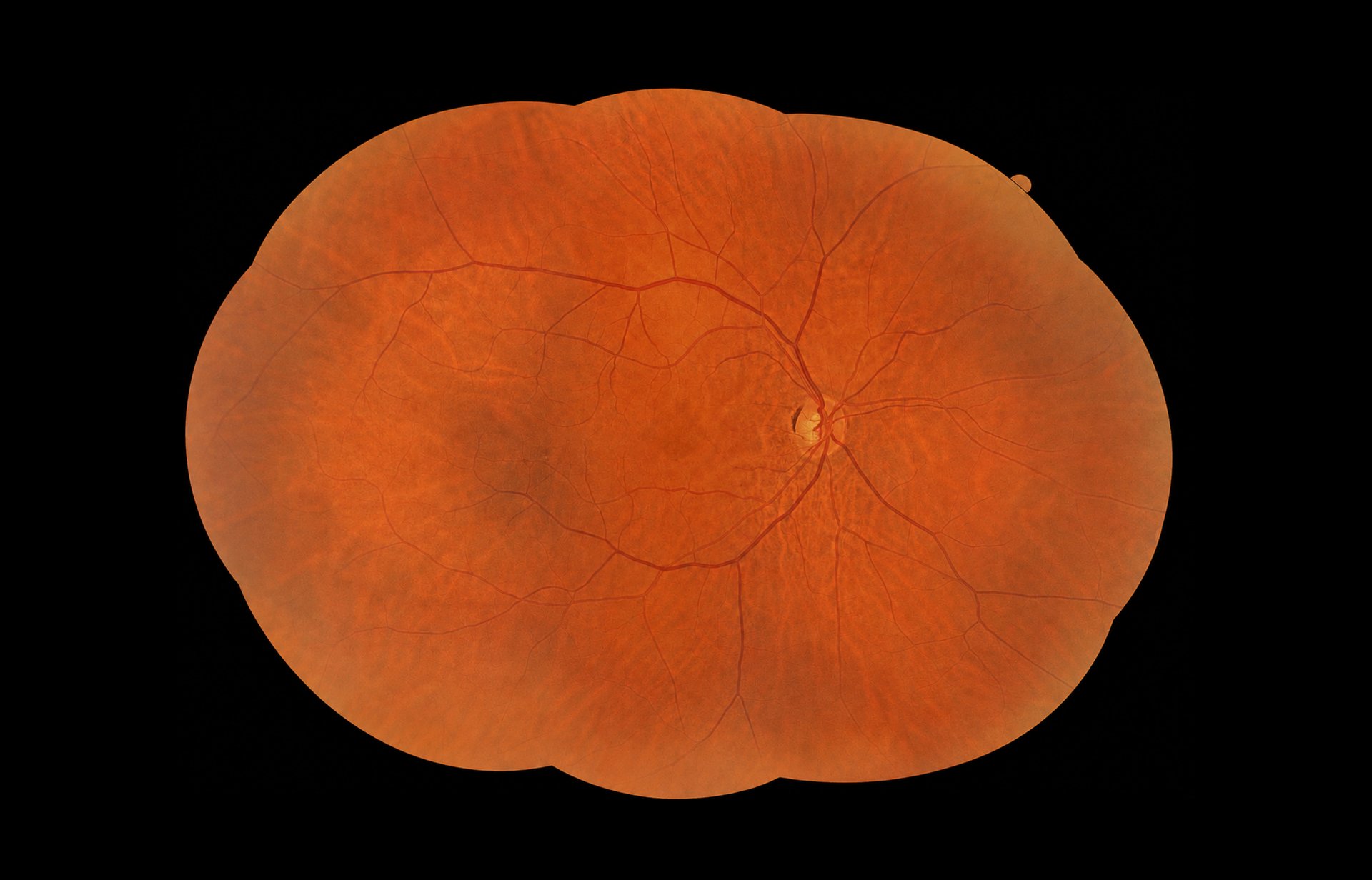
FUNDO provides retinal coverage of up to ~200° when measured from the center of the eye. Using up to nine fixation positions, the fully automated Mosaic mode captures peripheral retinal areas and creates panoramic true-color images without routine pupil dilation.
Five configurations are available:
- Horizontal Mosaic ×3,
- Vertical Mosaic ×3,
- Mosaic ×5,
- Mosaic ×9,
- Custom Mosaic ×2–9.
A unique binocular view enables side-by-side comparison of mosaic images from both eyes.
Technical Specifications
| Parameter | FUNDO |
|---|---|
| Type | Non-mydriatic automatic fundus camera |
| Photograph type | Color Fundus/Anterior, IR Fundus/Anterior, FAF – Green*, FAF – Blue*, FA*, ICGA* |
| Angular field of view (standard pupil) | 90° (60°) ± 5% |
| Angular field of view (small pupil) | 67° (45°) ± 5% |
| Mosaic field of view (standard pupil) | 198° (132°) ± 5%* |
| Minimum pupil size | 3.3 mm (small pupil) |
| Camera | 20.3 MP CMOS |
| Flash adjustment, gain, exposure | Auto, Manual |
| Intensity levels | High, Normal, Low |
| Focus adjustment range | -25 D to +25 D |
| Operating distance | 31 mm |
| Alignment method | Fully automatic, Automatic, Manual |
| Internal fixation target | OLED display with adjustable target shape, position, steady/blinking light mode, and blink rate |
| External fixation target | Yes |
| Dimensions (L×W×H) | 490 × 350 × 530 mm |
| Weight | 19 kg |
| Power supply | 100–240 V, 50/60 Hz |
| Power consumption | 90–110 VA |
| * Optional feature. Selectable during the ordering process. | |