COMING SOON
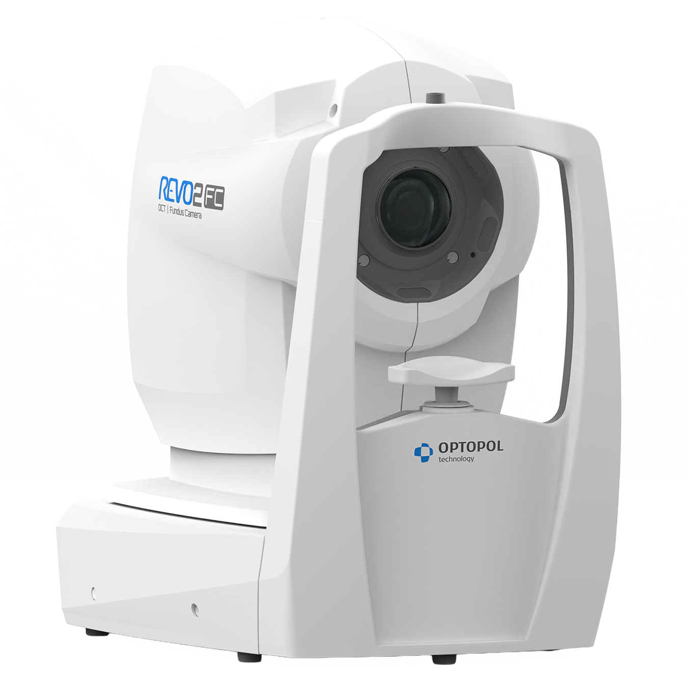
REVO 2
Widefield OCT & Fundus Imaging
One Platform. Endless Possibilities
REVO 2 represents the next generation of the proven REVO platform, bringing widefield retinal imaging to the forefront of ophthalmic diagnostics.
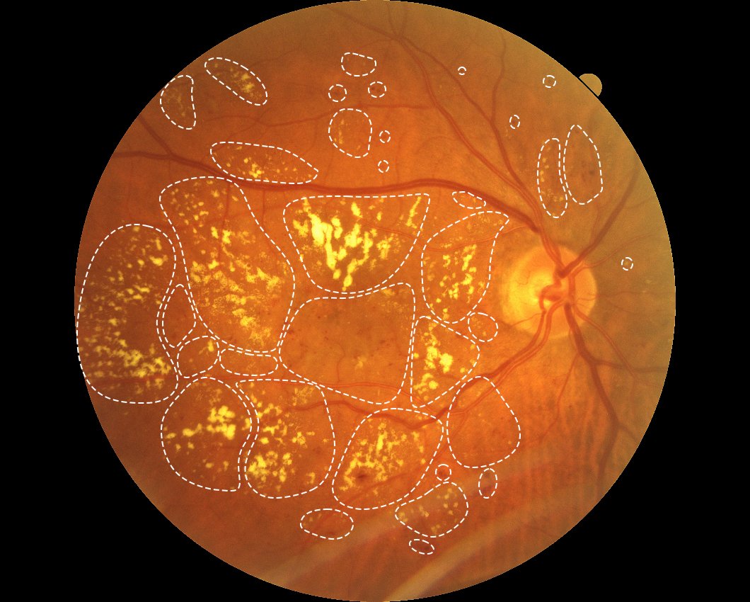
By combining widefield OCT imaging of up to approximately 115° with widefield color fundus imaging, the system enables wider visualization of central and peripheral retinal structures in a single examination. Building on the REVO All-in-One concept, the REVO 2 integrates a broad range of advanced fundus imaging modalities, including green and blue fundus autofluorescence (FAF), fluorescein angiography (FA), indocyanine green angiography (ICGA). Embedded AI technology supports both OCT and fundus imaging, it analyzes images, performs segmentation, detects anomalies, and supports result interpretation.
Create your dream all-in-one OCT
Multimodal Fundus Imaging
Choose the autofluorescence mode you need–or use both for a more complete assessment:
- Faf Green – Green Fundus Autofluorescence
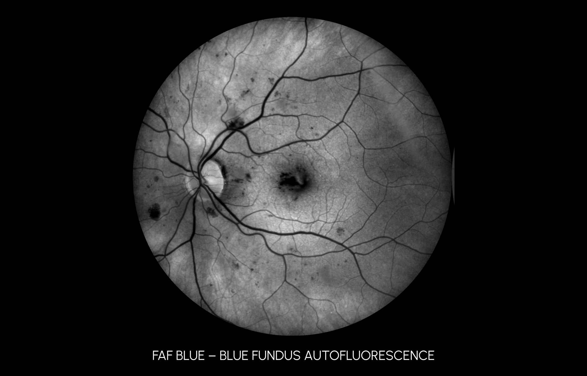
- Faf Blue – Blue Fundus Autofluorescence
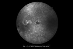
- Fa – Fluorescein Angiography
- Icga — Indocyanine Green Angiography
Ultra-Widefield Fundus Imaging
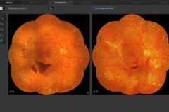
The REVO 2 enables ultra-widefield fundus imaging, providing retinal coverage of up to ~200° when measured from the spherical center of the eye and offering a broader perspective in fundus imaging
Widefield & Ultra-Widefield Imaging
23 mm Wide. 6 mm Deep. High-Resolution OCT.
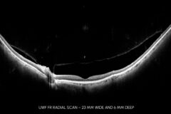
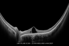
REVO 2 delivers widefield OCT imaging of up to 90°, combining broad retinal coverage with high-resolution visualization of retinal structures. With the optional ultra-widefield module, the field of view can be extended up to approximately 115° in a single scan.
Full Auto SD-OCT
Color Fundus Camera
OCT-A Angiography*
This non-invasive dye free technique enables the visualization of the microvasculature of the retina.
T-OCT™ Topography*
Detect corneal abnormalities with this pioneering way to capture detailed corneal curvature maps.
B-OCT® Biometry*
Perform automated measurements of the entire ocular structure and combine a biometer into your OCT.
IOL Calculation**
Keep your library up to date by calculating IOL implant parameters using the latest IOL database standard IOLCon.org.
* Available as an optional software module add-on function
** Requires B-OCT® Biometry module
Widefield Fundus Camera
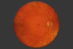
The integrated 20.3 MP fundus color camera captures detailed, high-quality retinal images with a wide view of the retina. Fully automated operation makes image acquisition fast, easy and consistent. Imaging is possible even through small pupils. The device automatically adjusts imaging parameters based on the IR fundus preview and automatically selects the appropriate flash intensity level for non-mydriatic examination of both eyes.
Fundus Analysis Tools
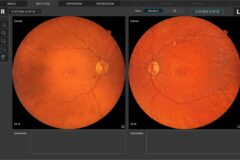
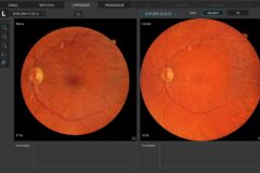
A comprehensive range of fundus views, analysis functions, and image-processing tools support efficient assessment, documentation, and follow-up of retinal conditions, enabling fast workflow in both routine clinical practice and retinal screening programs.
Available functions include:
- single-eye and bilateral imaging,
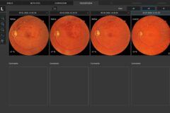
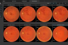
- chronological comparison of fundus photographs, including x4 and x8 progression views, for monitoring disease progression,
- image enhancement tools: Emboss+, Emboss-, Cobalt, Digital Cobalt, and Digital Red-Free,
- measurement functions for quantitative image assessment Area, Distance, and C/D,
- AI anomaly detection.
Multimodal Fundus Imaging
Choose the autofluorescence mode you need–or use both for a more complete assessment.
FAF Green – Green Fundus Autofluorescence
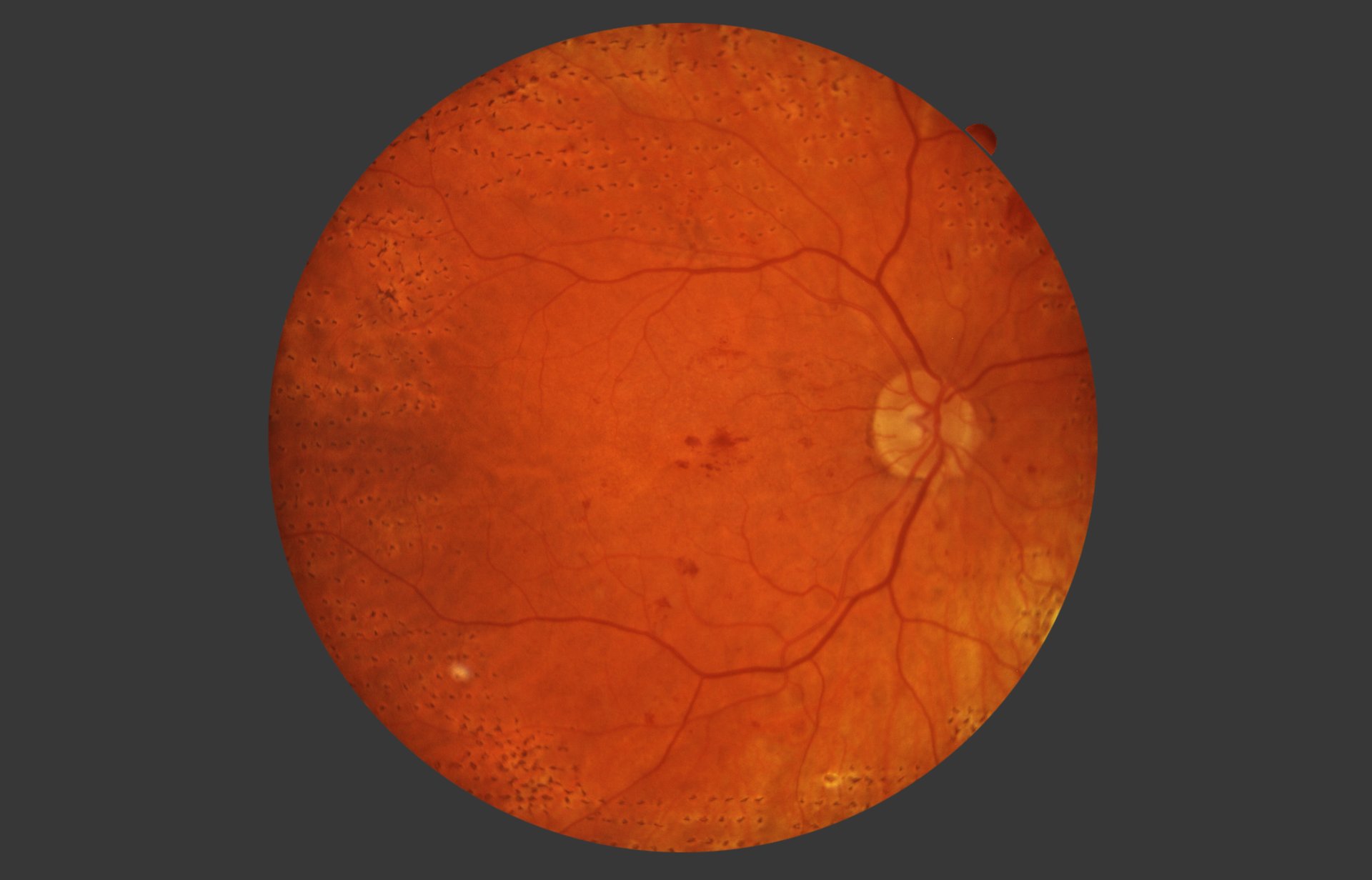
Green fundus autofluorescence supports the assessment of metabolic and structural changes in the retinal pigment epithelium (RPE) and outer retina, particularly in chorioretinal and degenerative retinal disorders.
FAF Blue – Blue Fundus Autofluorescence
Blue fundus autofluorescence provides complementary information on RPE integrity, lipofuscin-related changes, and outer retinal abnormalities.
Two angiography modes for deeper vascular insight—retinal with FA, choroidal with ICGA.
FA – Fluorescein Angiography
Fluorescein angiography enables the assessment of retinal vascular perfusion, leakage, microaneurysms, neovascularization, inflammatory vascular changes, and areas of capillary non-perfusion or ischemia.
ICGA — Indocyanine Green Angiography
Indocyanine green angiography supports the evaluation of choroidal circulation and vasculature, including polypoidal lesions, occult choroidal neovascularization, and deeper chorioretinal vascular abnormalities.
Ultra-Widefield Fundus Imaging
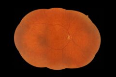
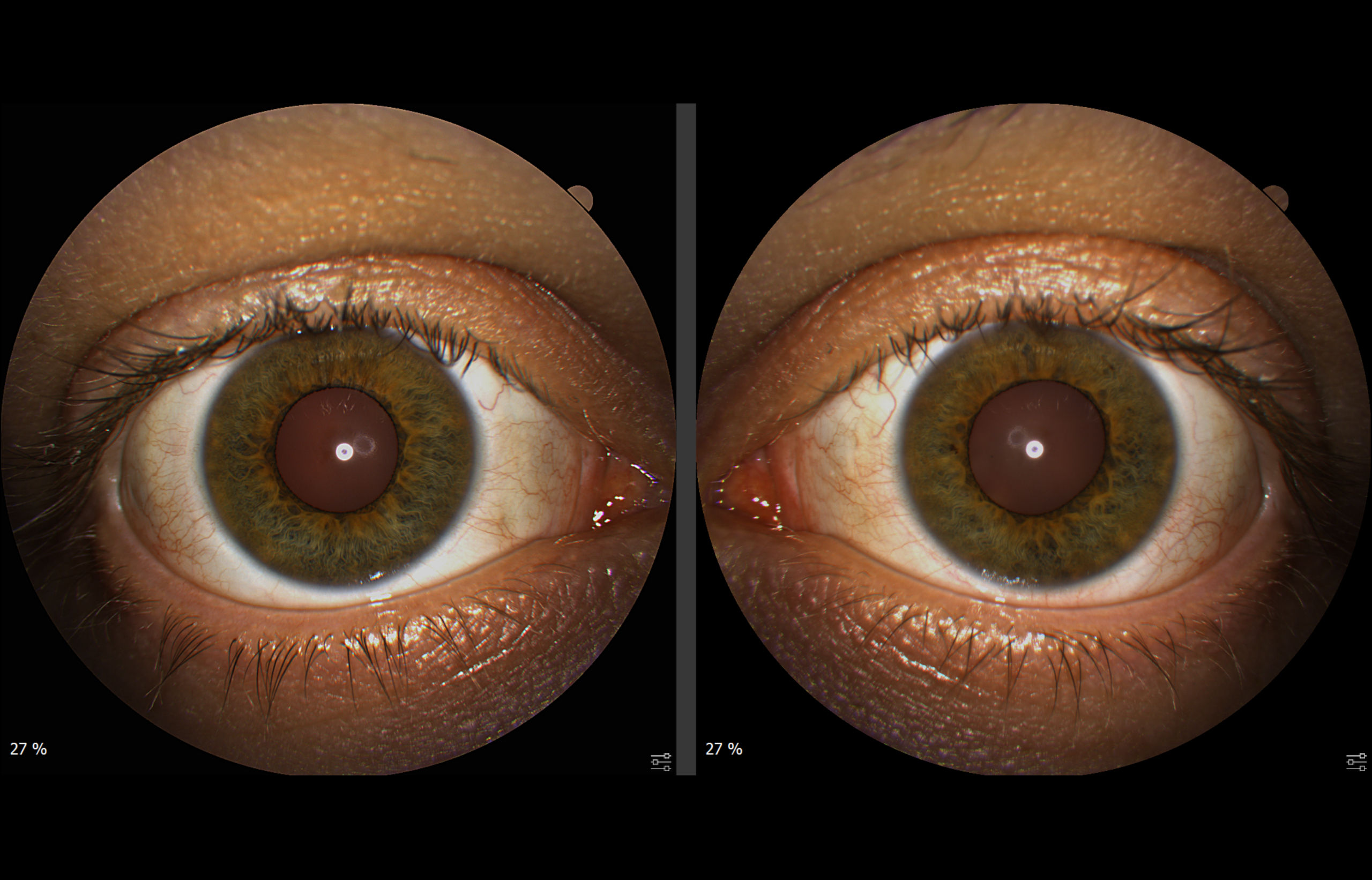
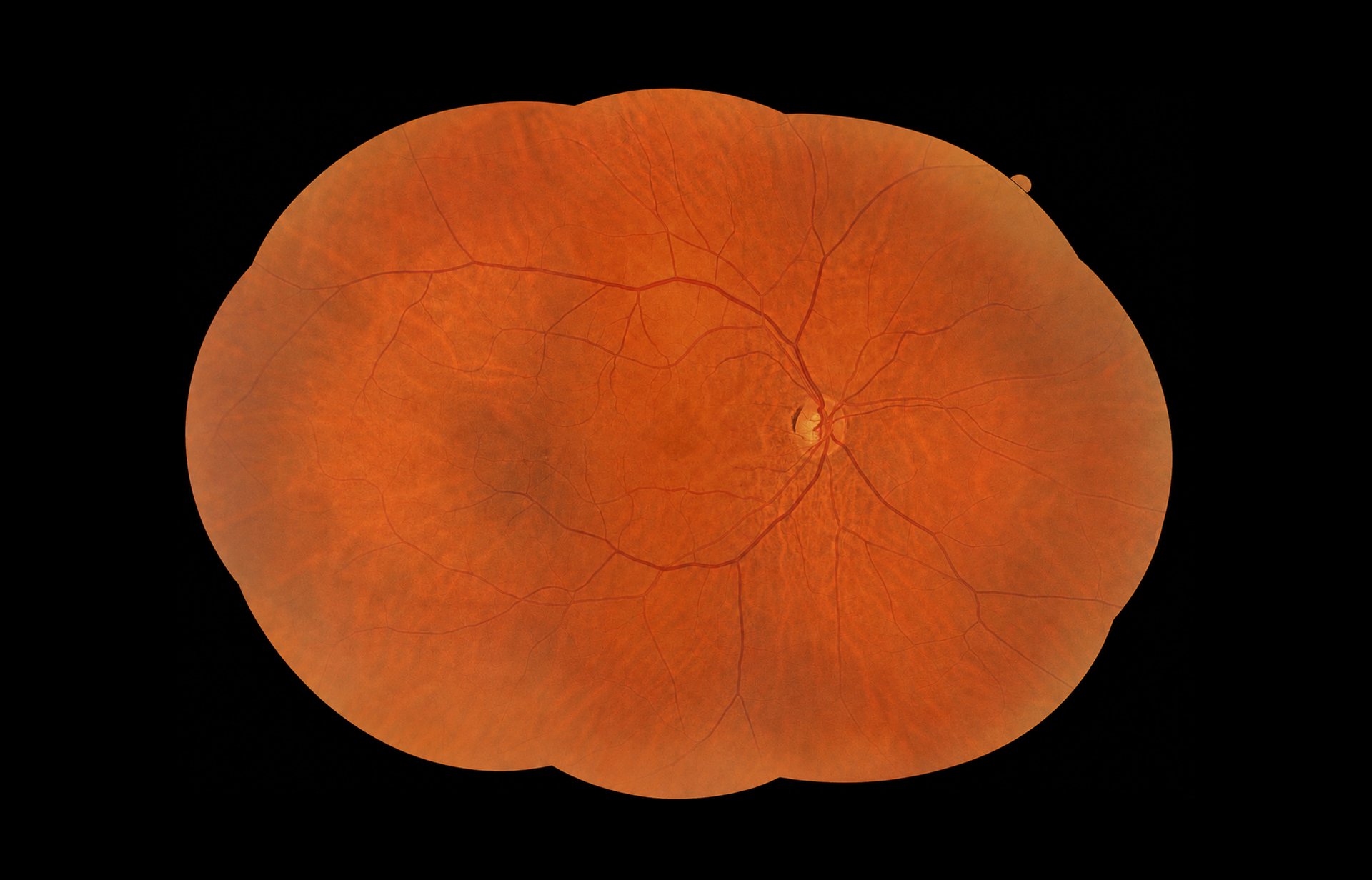
The REVO 2 enables ultra-widefield fundus imaging, providing retinal coverage of up to ~200° when measured from the spherical center of the eye and offering a broader perspective in fundus imaging. Mosaic Photo mode uses multiple internal fixation positions to capture peripheral retinal areas and automatically generate high-quality, true-color ultra-widefield mosaic images. Pupil dilation is not required.
Five configurations are available:
- Horizontal Mosaic ×3,
- Vertical Mosaic ×3,
- Mosaic ×5,
- Mosaic ×9,
- Custom Mosaic ×2–9.
Key features:
- fully automatic mosaic capture process,
- intuitive voice guidance for faster fixation shifts and image acquisition,
- simple operation and minimal training – select the configuration and press Start,
- instant mosaic generation during image acquisition,
- no delays caused by image transfer or by manual or automatic image superimposition.
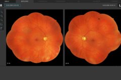
A unique binocular view enables side-by-side analysis of ultra-widefield images from both eyes and longitudinal comparison across visits.
Widefield & Ultra-Widefield Imaging
23 mm Wide. 6 mm Deep. High-Resolution OCT
REVO 2 delivers widefield OCT imaging of up to 90°, combining broad retinal coverage with high-resolution visualization of retinal structures. With the optional ultra-widefield module, the field of view can be extended up to approximately 115° in a single scan.
Ultra-widefield imaging captures both the macula and far retinal periphery, supporting the detection and assessment of peripheral changes in the posterior segment. It also provides 3D analysis, image averaging in Enhancement Mode, and Angio OCT for visualization of peripheral perfusion abnormalities.
Combined with Extended Depth™ software and Full Range scanning technology, the system extends imaging capabilities for deeper and more challenging examinations.
AI-Powered Analysis & Imaging
Clearer images. Smarter analysis. Faster, more confident decisions.
Embedded AI technology supports OCT and fundus imaging across the diagnostic workflow, helping improve image quality, automate analysis, and assist clinical interpretation.
AI Drusen Detection & Monitoring
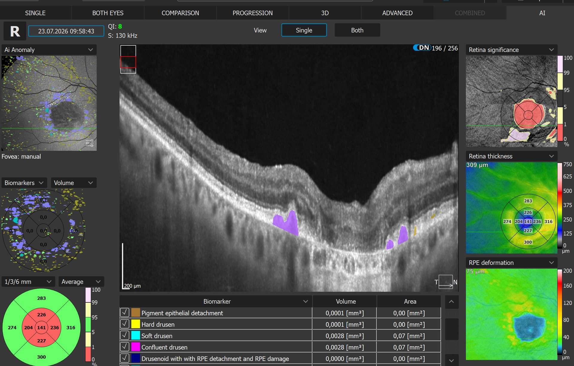
Automatic detection and quantification of different drusen types and associated RPE changes, including hard drusen, soft drusen, confluent drusen, pigment epithelial detachment, drusenoid with RPE detachment, and RPE damage.
Drusen are automatically detected and quantified by number, area, and volume, enabling detailed assessment during a single visit and longitudinal monitoring of changes across follow-up visits.
AI Segmentation
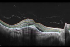
AI-based segmentation of the posterior segment, anterior chamber, and cornea enables more accurate detection of anatomical layer boundaries. This improves clinical evaluation, supports reliable assessment of retinal, anterior chamber, and corneal pathologies, and provides more precise quantification in routine examinations. Enhanced segmentation is particularly valuable in follow-up examinations, allowing clinicians to monitor changes over time more precisely, including in patients already under observation.
AI DeNoise
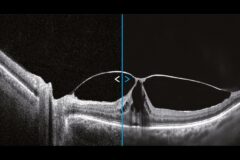
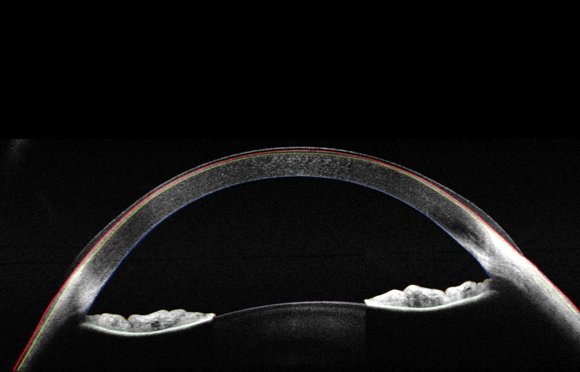
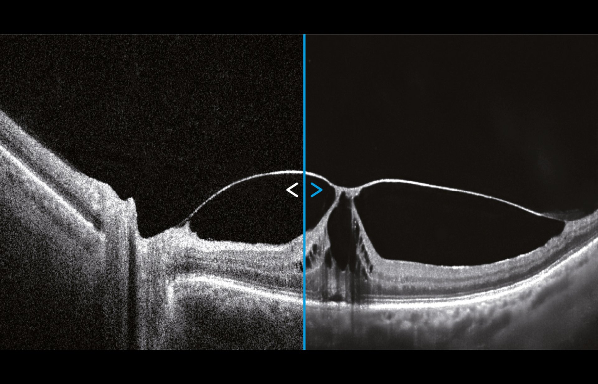
AI DeNoise uses artificial intelligence to enhance a single tomogram to a quality comparable to that of an averaged image obtained from multiple scans. It automatically reduces noise, producing clearer and smoother images.
The function is available for all tomograms and software views, including 3D.
Ultra-Widefield OCT Angiography
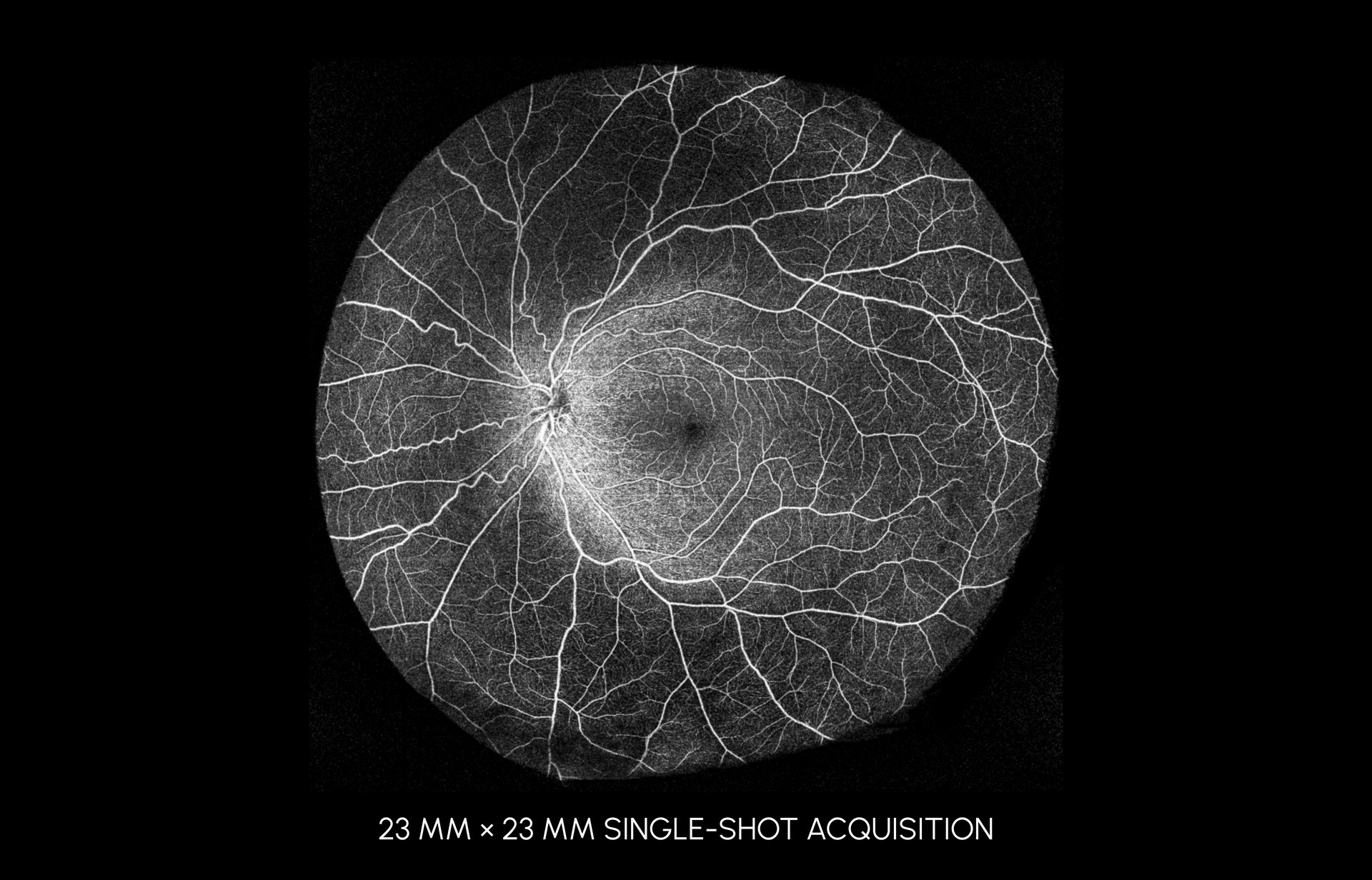
OCT Angiography is a non-invasive, dye-free technique for visualizing the retinal microvasculature, enabling detailed vascular imaging across a wide retinal field. Combined blood flow and structural imaging provide valuable diagnostic information for a wide range of retinal diseases.
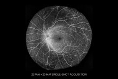
Angiography scans enable detailed assessment of the vascular structure of the macula, retinal periphery, and optic disc. With acquisition times of just 1.6 seconds in standard resolution and 3 seconds in high resolution, OCT Angiography can be easily incorporated into routine diagnostic practice. A comprehensive set of analysis views allows clinicians to observe, monitor, and compare changes in the retinal microvasculature of both eyes.
Anterior Chamber Imaging & Angle Analysis
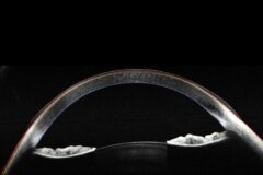
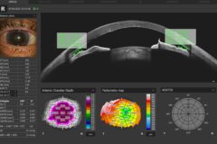
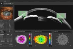
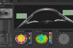
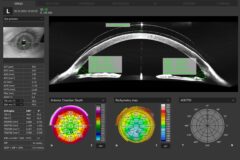
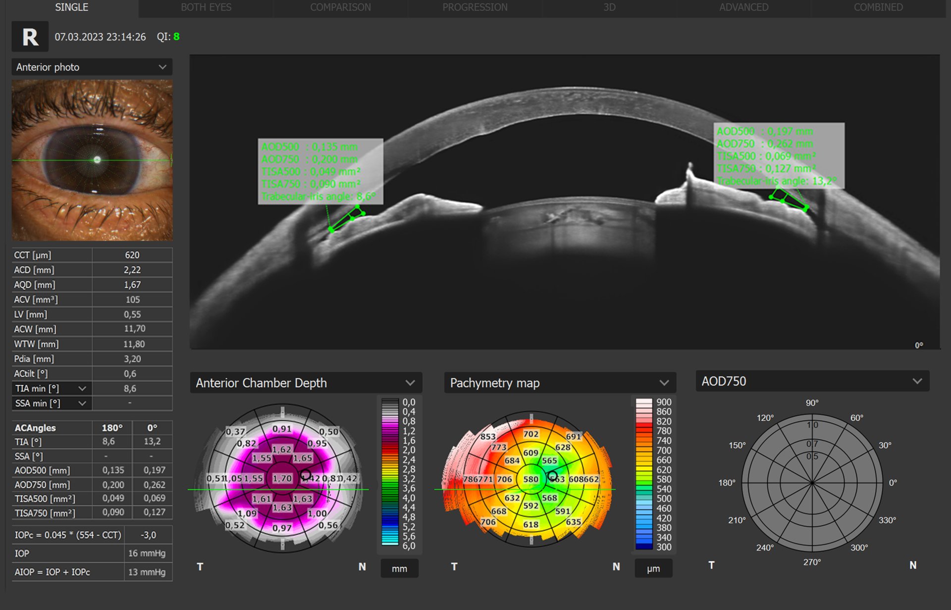
Integrated anterior segment optics enable imaging without the need for additional lenses or a forehead adapter. The operator can visualize and quantify the entire anterior segment or focus on a selected area for detailed evaluation.
Automated anterior chamber analysis enables fast and convenient assessment, providing a workflow experience comparable to that of dedicated anterior segment analysis devices, such as AS-OCT and Scheimpflug systems.
OCT gonioscopy enables visualization, measurement, and mapping of both iridocorneal angles and iris configuration in a single high-resolution 360° radial scan, supporting comprehensive glaucoma assessment.
Automated analysis provides anterior chamber parameters, including CCT, ACD, AQD, ACV, LV, ACW, WTW, Pdia, and AC tilt. It also provides semi-automated angle analysis with parameters such as TIA, SSA, AOD500, AOD750, TISA500, and TISA750, as well as pachymetry, anterior chamber depth, stromal thickness, and epithelial thickness maps.
Technical Specifications
| Parameter | REVO 2 FC130 | REVO 2 FC |
|---|---|---|
| OCT | ||
| Technology | Spectral-Domain OCT | |
| Scanning speed | 130 000 A-scans/s | 80 000 A-scans/s |
| Axial resolution | In tissue 5.5 μm, Digital 2.8 μm | |
| Light source | SLED | |
| Central wavelength | 850 nm | |
| Bandwidth | 50 nm | |
| Lateral range | Posterior (3 to 20) mm ± 5%, Angio OCT (3 to 20) mm ± 5%, Anterior (3 to 18) mm ± 5% | |
| Lateral resolution | 12 μm | |
| Axial range | Standard 2.8 mm, Full Range ~6.0 mm | |
| Minimum pupil size (OCT) | 2.0 mm | |
| Focus adjustment range | -25 D to +25 D | |
| Fundus tracking | Live / Interactive – AccuTrack active real time, iTracking | |
| Operating distance | 31 mm | |
| Alignment method | Fully automatic, Automatic, Manual | |
| Glaucoma analysis | RNFL, ONH morphology, DDLS, OU/Hemisphere asymmetry, Ganglion analysis (RNFL+GCL+IPL, GCL+IPL), Structure + Function² | |
| Retinal analysis | Retinal thickness, Inner Retinal thickness, Outer Retinal thickness, RNFL+GCL+IPL thickness, GCL+IPL thickness, RNFL thickness, RPE deformation, MZ/EZ-RPE thickness | |
| Corneal Topography Map¹ | Axial maps (Anterior, Posterior), Refractive Power (Kerato, Anterior, Posterior, Total), Net Map, Axial True Net, Equivalent Keratometer, Elevation maps (Anterior, Posterior), Height maps, KPI (Keratoconus Prediction Index) | |
| Biometry¹ | AL, CCT, ACD, LT, P, WTW | |
| IOL Calculator³ | Hoffer Q, Holladay I, Haigis, Theoretical T, Regression II | |
| Anterior (no adapter required) | Pachymetry map, Epithelium map, Stroma map, Anterior Chamber Depth map, Anterior Chamber Parameters: CCT, ACD, AQD, ACV, LV, ACW, WTW, Pdia, AC tilt, AOD 500/750 map, TISA 500/750 map | |
| Angiography OCT¹ | Vitreous, Retina, Choroid, Superficial Plexus, Deep Plexus, Outer Retina, Choriocapillaris, RPCP, SVC, DVC, ICP, DCP, Custom segmentation, Enface imaging, FAZ, VFA, NFA, Vessel Area Density quantification, Skeleton Area Density quantification, Thickness maps | |
| Angiography mosaic | Auto Mosaic modes: 10×10, 10×6, 12×5, 7×7 Manual up to 12 images |
Auto Mosaic modes: 10×6 Manual up to 12 images |
| Voice support | Yes | |
| Internal fixation target | OLED display (target shape, position, steady/blinking light, and blinking speed can be changed) | |
| External fixation target | Yes | |
| Dimensions (L×W×H) / Weight | 490 × 350 × 530 mm / 30 kg | |
| Power supply | 100–240 V, 50/60 Hz | |
| Power consumption | 90–110 VA | |
| UWF LENS⁴ | ||
| UWF scan angle | ~115°⁵ ± 5% | |
| UWF working distance | 16 mm | |
| UWF scan types | 3D, Radial, Line, Angio¹, Full Range Radial, Full Range Line | |
| UWF fundus tracking | iTracking | |
| UWF lateral range | Standard 2.8 mm, Full range ~6 mm | |
| FUNDUS CAMERA | ||
| Type | Non-mydriatic automatic fundus camera | |
| Photography mode | Color Fundus/Anterior, IR Fundus/Anterior, FAF – Green*, FAF – Blue*, FA*, ICGA* | |
| Angular field of view (standard pupil) | 90° (60°) ± 5% | |
| Angular field of view (small pupil) | 67° (45°) ± 5% | |
| Mosaic field of view (standard pupil) | 198° (132°) ± 5% | |
| Resolving power on fundus camera | Center: 60 lp/mm or more, Middle (r/2): 40 lp/mm or more, Periphery (r): 25 lp/mm or more | |
| Minimum pupil size | Small pupil 3.3 mm | |
| Camera | Built-in 20.3-megapixel CMOS camera | |
| Flash adjustment, gain, exposure | Auto / Manual | |
| Intensity levels | High, Normal, Low | |
| * Optional feature, selectable at the time of order ¹ Optional software module ² Via connection with PTS software version 3.8 or higher ³ The Biometry module and a separate license for the IOL Calculator are required ⁴ Ultra-Widefield imaging is available with the optional UWF lens ⁵ Measured from the center of the eye |
||